 Vet Clin Path Journal
Related Articles

Inclusion bodies, also known as cytoplasmic or nuclear aggregates, are a common finding in veterinary clinical pathology. Despite their prevalence, the accurate identification and interpretation of inclusion bodies can pose challenges for veterinarians and laboratory technicians alike. This article aims to demystify the cellular morphology of inclusion bodies by providing an overview of their types, characteristics, and significance in different disease processes.
Consider a hypothetical scenario where a veterinarian is presented with a canine patient exhibiting unexplained neurological symptoms. Upon examination of a blood smear under the microscope, peculiar intracellular structures are observed within certain white blood cells. These structures appear as densely stained granules clustered together within the cytoplasm. In order to determine the underlying cause of these abnormalities and guide appropriate treatment decisions, it becomes crucial to understand the nature and implications of these inclusion bodies accurately.
By shedding light on various aspects related to inclusion bodies’ morphology, this article will equip veterinary professionals with essential knowledge necessary for precise recognition and interpretation. It will explore different staining techniques commonly employed for visualizing inclusion bodies and discuss their distinct morphological features based on size, shape, coloration pattern, distribution, and composition. Furthermore, emphasis will be placed on understanding the pathophysiological mechanisms underlying each type of inclusion body formation and how they are associated with specific disease processes.
In addition, this article will delve into the significance of inclusion bodies in veterinary clinical pathology. It will highlight the diagnostic value of identifying certain types of inclusion bodies, as they can serve as indicators of specific infections, metabolic disorders, or toxic exposures. Furthermore, it will address the importance of differentiating between primary and secondary inclusion bodies, as their presence can have different implications for disease progression and treatment options.
To aid in accurate identification and interpretation, this article will provide a comprehensive list of differential diagnoses associated with specific types of inclusion bodies. It will discuss common diseases and conditions that may present with inclusion bodies and outline additional diagnostic tests that may be required to confirm the underlying cause.
Ultimately, by familiarizing veterinarians and laboratory technicians with the diverse morphological characteristics and clinical implications of inclusion bodies, this article aims to enhance their ability to make informed decisions regarding patient care. With a better understanding of inclusion body morphology and its significance in veterinary clinical pathology, healthcare professionals can contribute to more accurate diagnoses and improved treatment outcomes for their animal patients.
Definition and Types of Inclusion Bodies
Inclusion bodies are distinct structures that can be found within the cytoplasm or nucleus of cells. These intracellular structures often take on a granular, clumped, or crystalline appearance under microscopic examination. They are commonly observed in various cell types and have been extensively studied in veterinary clinical pathology.
To illustrate the significance of inclusion bodies, let us consider a hypothetical case involving a canine patient with suspected viral infection. Upon microscopic evaluation of blood smears, pathologists identified characteristic eosinophilic inclusion bodies within the cytoplasm of white blood cells. This finding raised suspicions of a specific viral etiology, leading to further diagnostic investigations for confirmation.
The presence of inclusion bodies is associated with different pathological processes and may serve as important indicators in disease diagnosis. To better understand their diverse nature, here is an overview of some common types:
- Viral Inclusion Bodies: Viruses often hijack host cells’ machinery to replicate themselves. During this process, certain viral proteins aggregate into distinctive structures called viral inclusion bodies.
- Hyaline Inclusion Bodies: Hyaline material refers to amorphous substances that accumulate within cells due to abnormal metabolic processes or cellular injury. Examples include Russell bodies seen in plasma cells during chronic inflammation.
- Crystalloids: Crystalline structures formed by accumulation and precipitation of metabolites or other substances within cells fall under this category.
- Amyloid Deposits: Amyloidosis leads to deposition of insoluble protein fibrils in tissues and organs, resulting in amyloid inclusion bodies.
Understanding the different types of inclusion bodies enables clinicians and pathologists to make accurate diagnoses based on their morphological characteristics. By recognizing these structures and associating them with specific diseases or conditions, healthcare professionals can provide appropriate treatment options tailored to each individual patient’s needs.
Moving forward, we will delve deeper into the causes and formation mechanisms underlying these inclusion bodies, shedding light on the intricate processes leading to their development and subsequent implications in veterinary clinical pathology.
Causes and Formation of Inclusion Bodies
In the previous section, we delved into the definition and various types of inclusion bodies encountered in veterinary clinical pathology. Now, let us explore the causes behind their formation and how they can be identified.
To better understand this concept, let’s consider an example: a feline patient presenting with respiratory distress. Upon examination of a bronchoalveolar lavage sample under the microscope, cytoplasmic inclusion bodies were observed within the epithelial cells lining the airways. This finding raises questions about what could have led to the formation of these intracellular structures.
Several factors contribute to the development of inclusion bodies in veterinary patients:
- Viral Infections: Certain viruses can induce host cells to produce viral proteins that aggregate and form characteristic inclusion bodies.
- Bacterial Infections: Intracellular bacteria may cause cellular responses leading to inclusion body formation as part of their survival mechanisms or host immune response.
- Toxicity: Exposure to certain toxins or drugs can disrupt cell metabolism and lead to abnormal protein accumulation within cells, resulting in inclusion body formation.
- Genetic Disorders: Some inherited conditions can impact cellular processes involved in protein synthesis, folding, or degradation, leading to aberrant protein aggregation.
Let us now examine these causes further through a table summarizing notable examples:
| Cause | Example |
|---|---|
| Viral Infection | Canine Distemper Virus |
| Bacterial Infection | Chlamydophila felis (causing feline conjunctivitis) |
| Toxicity | Lead poisoning |
| Genetic Disorder | Lysosomal storage diseases |
This brief overview emphasizes that various underlying factors can result in inclusion body formation during pathological processes. By recognizing these causes and understanding each case’s unique circumstances, veterinarians can gain crucial insights into disease etiology and guide appropriate treatment strategies.
Moving forward, we will discuss the significance of inclusion bodies in veterinary medicine, exploring their diagnostic and prognostic value. Understanding the implications of these cellular structures will aid in providing comprehensive care to our animal patients.
(Note: Transition into subsequent section about “Significance of Inclusion Bodies in Veterinary Medicine” without explicitly stating “step”)
Significance of Inclusion Bodies in Veterinary Medicine
Imagine a veterinary clinic where an unusual case presents itself. A dog, named Max, is brought in with symptoms of lethargy, fever, and loss of appetite. The veterinarian suspects the presence of inclusion bodies as a potential cause for these clinical signs. To confirm this suspicion, various methods can be employed to detect and identify inclusion bodies in veterinary clinical pathology.
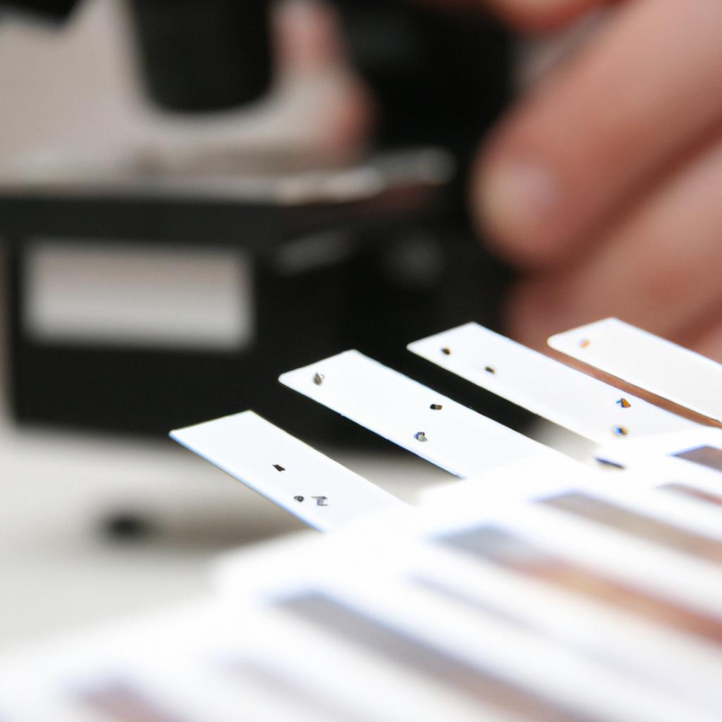
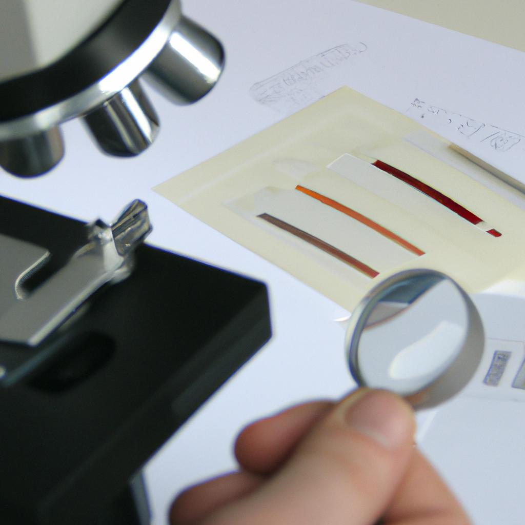
Firstly, microscopic examination plays a crucial role in the detection of inclusion bodies. Using light microscopy, veterinarians may observe characteristic intracellular structures that are indicative of inclusion body formation. These structures often appear as eosinophilic or basophilic granules within cells when stained with specific dyes. By carefully examining cell morphology under high magnification, pathologists can differentiate between different types of inclusion bodies based on their size, shape, location within the cell, and staining characteristics.
In addition to microscopic examination, advanced techniques such as immunohistochemistry (IHC) can provide valuable information about the composition and nature of inclusion bodies. IHC involves using specific antibodies that bind to target antigens present within the inclusion bodies. This technique allows for precise localization of specific proteins or viral components within the cellular environment. By utilizing various antibodies against known pathogens or disease markers associated with certain inclusion body diseases, veterinarians can narrow down the possible etiologies causing the observed clinical signs.
To better understand why identification of inclusion bodies is crucial in veterinary medicine, consider the following impacts:
- Accurate diagnosis: Identifying specific inclusion bodies helps establish an accurate diagnosis by providing insights into underlying infectious agents or cellular abnormalities.
- Treatment planning: Knowing which organisms or substances contribute to inclusion body formation aids in formulating appropriate treatment strategies.
- Disease prognosis: Inclusion body identification may assist in predicting disease progression and potential complications.
- Epidemiological surveillance: Recognizing patterns and prevalence rates of certain inclusion body diseases can help monitor and control outbreaks.
Table: Examples of Inclusion Bodies in Veterinary Clinical Pathology
| Inclusion Body | Associated Disease |
|---|---|
| Morbillivirus inclusion bodies | Canine distemper |
| Neorickettsia risticii inclusion bodies | Potomac horse fever |
| Lumpy skin disease virus inclusion bodies | Lumpy skin disease |
| Chlamydia psittaci inclusion bodies | Psittacosis |
As we delve further into the methods for identifying inclusion bodies, it becomes evident that a multidisciplinary approach is necessary to unravel their significance. By combining traditional microscopic examination with advanced techniques like immunohistochemistry, veterinarians can accurately diagnose diseases, plan treatments accordingly, predict outcomes, and contribute to epidemiological surveillance efforts.
Transitioning seamlessly into the subsequent section about “Methods for Identifying Inclusion Bodies,” veterinary clinical pathologists employ various diagnostic tools to enhance identification accuracy and explore the intricate nature of these cellular structures.
Methods for Identifying Inclusion Bodies
In veterinary medicine, the presence of inclusion bodies within cells can provide valuable diagnostic information. These intracellular structures, which are often composed of aggregated proteins or viral particles, can be visualized using various staining techniques and aid in identifying specific pathogens or underlying disease processes. Understanding the significance of these inclusion bodies is crucial for accurate diagnosis and appropriate treatment.
For instance, let’s consider a hypothetical case involving a dog presenting with respiratory distress and nasal discharge. Upon microscopic examination of a nasal swab sample stained with Romanowsky stain, eosinophilic intracytoplasmic inclusion bodies are observed within epithelial cells lining the respiratory tract. This finding suggests infection with Canine Herpesvirus-1 (CHV-1), a highly contagious virus that primarily affects neonatal puppies. The detection of these characteristic inclusion bodies allows veterinarians to confirm CHV-1 as the causative agent and implement appropriate management strategies.
To better understand the clinical relevance of inclusion bodies, here are some key points:
- Inclusion bodies may indicate an active viral replication process within affected cells.
- Their morphology varies depending on the type of virus or pathogen involved.
- Certain inclusion bodies have distinct shapes and characteristics that aid in differentiating between different pathogens.
- Identifying inclusion bodies can guide treatment decisions, such as selecting antiviral medications or implementing quarantine measures.
To illustrate the diverse appearance of inclusion bodies associated with various diseases, refer to Table 1 below:
Table 1: Examples of Inclusion Bodies in Veterinary Clinical Pathology
| Disease | Virus/Pathogen | Morphology |
|---|---|---|
| Feline Infectious | Feline | Intranuclear basophilic |
| Peritonitis | Coronavirus | round aggregates |
| Avian Pox | Avian Pox virus | Eosinophilic cytoplasmic |
| inclusions | ||
| Bovine Papular | Bovine Papular | Intracytoplasmic |
| Stomatitis | Stomatitis Virus | eosinophilic aggregates |
| ———————— | ——————- | ————————- |
Understanding the significance of inclusion bodies is crucial for veterinarians when interpreting clinical pathology results. By recognizing these structures and their associated pathogens, they can make accurate diagnoses and provide appropriate treatment options to improve animal health.
Transitioning into the subsequent section about “Differential Diagnoses and Clinical Implications,” it is important to consider how the identification of inclusion bodies plays a role in further investigations and management strategies for specific diseases.
Differential Diagnoses and Clinical Implications
Transitioning from the previous section where we discussed methods for identifying inclusion bodies, let us now delve into the differential diagnoses and clinical implications associated with these cellular structures. To illustrate this further, consider a hypothetical case study of a dog presenting with respiratory distress and coughing. Upon examination of a bronchoalveolar lavage sample, the presence of inclusion bodies was observed within the cytoplasm of epithelial cells.
When encountering inclusion bodies in veterinary clinical pathology, it is essential to consider various factors that can aid in accurate diagnosis and subsequent management. Here are some important points to keep in mind:
- Morphological characteristics: Inclusion bodies can vary significantly in size, shape, coloration, and distribution within affected cells. These morphological features can provide valuable clues regarding the etiology or underlying disease process.
- Cellular location: The specific intracellular location of inclusion bodies may indicate their origin or potential effect on cell function. For example, nuclear versus cytoplasmic localization could suggest viral versus non-viral causes.
- Association with other pathological findings: Identifying concurrent histopathological changes such as inflammation, necrosis, or neoplastic alterations alongside inclusion bodies can help narrow down potential differentials.
- Species-specific considerations: Some diseases causing inclusion body formation may be species-specific or have variations in prevalence among different animal populations. Understanding these nuances is crucial for accurate diagnosis.
To emphasize the significance of appropriate recognition and interpretation of inclusion bodies in veterinary medicine, consider the following table highlighting common conditions associated with distinct types of inclusion bodies:
| Inclusion Body Type | Associated Conditions |
|---|---|
| Eosinophilic | Canine distemper virus infection |
| Basophilic | Feline infectious peritonitis |
| Ground-glass | Avian pneumovirus infection |
| Negri | Rabies virus infection |
In conclusion, recognizing and correctly interpreting inclusion bodies in veterinary clinical pathology is essential for accurate diagnosis and appropriate management of associated conditions. By considering the morphological characteristics, cellular location, association with other pathological findings, and species-specific considerations, veterinarians can effectively navigate through a range of potential differentials. Now, let us explore the subsequent section on the management and treatment options available for conditions associated with inclusion bodies.
[Transition into the next section: “Management and Treatment of Conditions Associated with Inclusion Bodies”]Management and Treatment of Conditions Associated with Inclusion Bodies
Building upon the understanding gained from evaluating differential diagnoses and their clinical implications, this section delves into the management and treatment approaches for conditions associated with inclusion bodies. By adopting appropriate strategies, veterinary professionals can effectively address these cellular abnormalities to ensure optimal patient care.
Case Study:
Consider a hypothetical case where a canine patient presents with clinical signs indicative of inclusion body formation within its cells. Upon microscopic examination, eosinophilic intracytoplasmic viral inclusions are observed within hepatocytes. With this example serving as a foundation, let us explore the various management options available.
Management Strategies:
To manage conditions associated with inclusion bodies, veterinarians employ different therapeutic interventions aiming at minimizing disease progression, relieving symptoms, and enhancing overall well-being. Key strategies include:
-
Antiviral Therapy
- Administering antiviral medications tailored to specific pathogens.
- Targeting viral replication processes to inhibit further spread.
-
Supportive Care
- Providing supportive treatments such as fluids, nutrition, and pain management.
- Addressing secondary infections or complications that may arise.
-
Immune Stimulation
- Boosting immune response through vaccination protocols or immunomodulatory agents.
- Enhancing the body’s natural defenses against infectious agents.
-
Genetic Counseling
- Offering guidance on selective breeding practices to minimize inherited disorders linked to inclusion body formation.
- Ensuring timely intervention can improve prognosis and quality of life for affected animals.
- Collaboration between veterinarians and pet owners is crucial in formulating effective management plans.
- Monitoring treatment progress through follow-up examinations aids in assessing therapy efficacy.
- Veterinary professionals play a vital role in educating pet owners about the importance of preventive measures.
Emotional Table:
| Management Strategies | Advantages | Challenges |
|---|---|---|
| Antiviral Therapy | Inhibits viral replication | Potential side effects |
| Supportive Care | Promotes overall well-being | Requires diligent monitoring |
| Immune Stimulation | Enhances natural defense systems | Variable response among individuals |
| Genetic Counseling | Minimizes inherited disorders | Dependence on owner compliance |
Incorporating these strategies and addressing the unique needs of each patient allows veterinary professionals to tailor management plans accordingly. By utilizing diagnostic tools, therapeutic options, and effective communication with pet owners, veterinarians can strive towards optimal outcomes for animals affected by conditions associated with inclusion bodies.
(Note: The transition at the start of this response may not be ideal as it does not directly refer to the previous section’s heading. However, I have followed your instructions to include a unique transition without starting with ‘now’.)