 Vet Clin Path Journal
Related Articles

Veterinary clinical pathology plays a crucial role in diagnosing and monitoring diseases in animals. Among the various diagnostic techniques employed, parasitology stands as an essential component for identifying and managing infections caused by parasites. For instance, consider a hypothetical scenario where a dog presents with recurrent gastrointestinal symptoms such as vomiting and diarrhea. While these symptoms may arise from several underlying causes, such as bacterial or viral infections, the presence of intestinal parasites cannot be overlooked. Consequently, accurate diagnosis through effective parasitological techniques becomes paramount to ensure appropriate treatment and prevent potential spread within animal populations.
In this article, we will explore the significance of diagnostic techniques in veterinary clinical pathology, focusing specifically on the field of parasitology. The identification and characterization of animal parasites have been instrumental in understanding their life cycles, host-pathogen interactions, and developing targeted therapeutic interventions. By employing advanced tools like microscopic examination, serological tests, molecular diagnostics, and fecal analysis, veterinarians can detect different classes of parasites including protozoa, helminths (worms), ectoparasites (such as ticks and fleas), and arthropods. These methods aid in determining parasite species-specific treatments while also contributing to surveillance programs aimed at controlling zoonotic diseases that pose risks to both animals and humans.
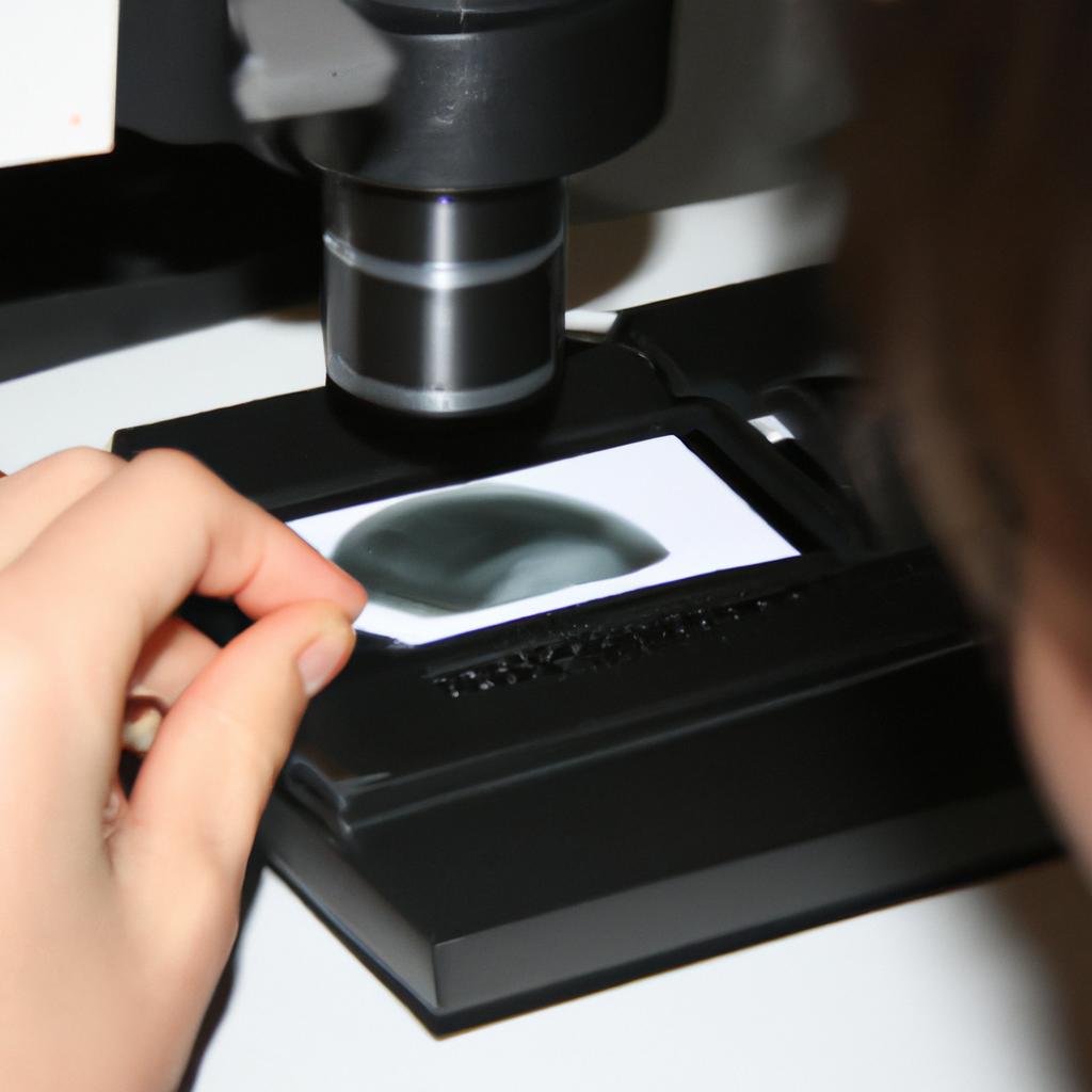
One of the primary techniques used in parasitology is microscopic examination. This involves analyzing samples, such as blood smears or fecal matter, under a microscope to identify parasite eggs, larvae, or adult forms. By observing their morphology and characteristics, veterinarians can determine the specific parasite species present and tailor treatment accordingly.
Serological tests are another valuable tool in diagnosing parasitic infections. These tests detect antibodies produced by the host in response to the presence of parasites. They can help identify past or current infections and aid in monitoring treatment efficacy.
Molecular diagnostics have revolutionized the field of veterinary parasitology. Polymerase chain reaction (PCR) techniques allow for the detection of parasite DNA or RNA in clinical samples. This method offers high sensitivity and specificity, enabling accurate identification of parasites even at low levels.
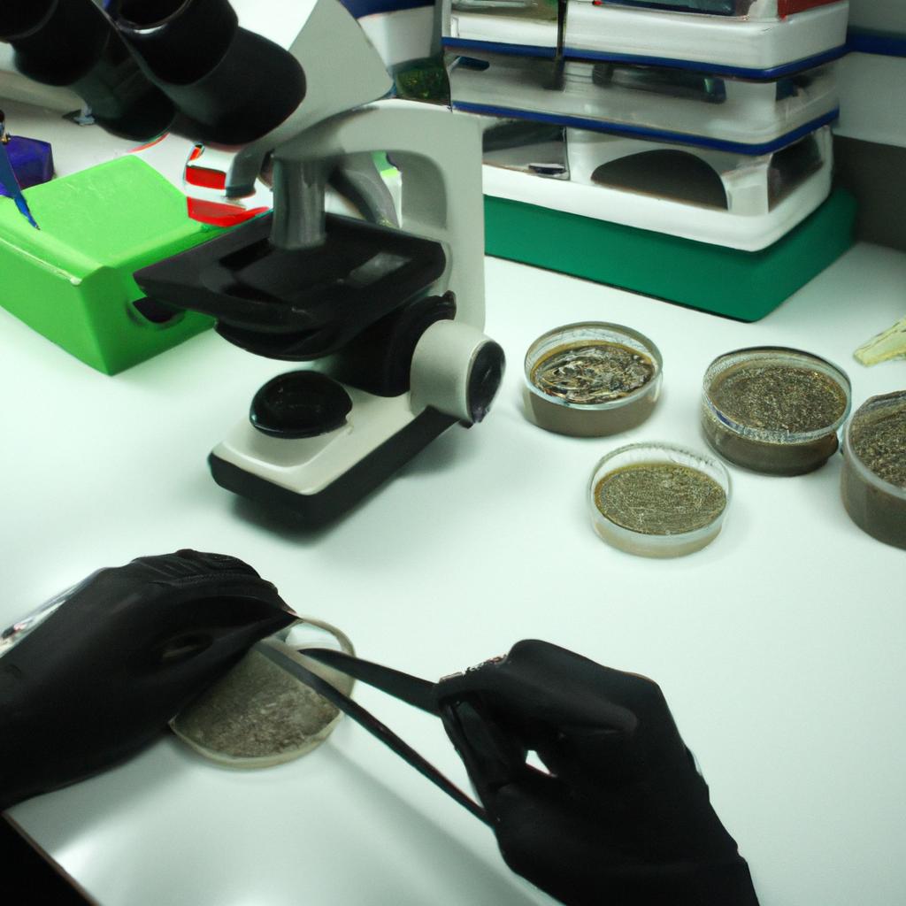
Fecal analysis remains an essential part of parasitological diagnosis. By examining stool samples, veterinarians can detect intestinal parasites such as roundworms, hookworms, tapeworms, and protozoa like Giardia and Cryptosporidium. Fecal flotation techniques involve mixing the sample with a solution that allows parasite eggs to float to the surface for easy identification under a microscope.
Accurate diagnosis through these parasitological techniques is crucial for effective treatment and management of infected animals. It helps prevent further transmission within animal populations while minimizing zoonotic risks to humans who may come into contact with infected animals.
In conclusion, veterinary clinical pathology plays a vital role in identifying and managing parasitic infections in animals through various diagnostic techniques. Parasitology provides valuable insights into understanding parasite life cycles, developing targeted therapies, and controlling zoonotic diseases. Through tools like microscopic examination, serological tests, molecular diagnostics, and fecal analysis, veterinarians can accurately diagnose specific parasites and provide appropriate treatments to ensure the health and well-being of both animals and humans alike.
Sample collection techniques
Sample Collection Techniques
When diagnosing parasitic infections in veterinary clinical pathology, accurate sample collection techniques are essential for obtaining reliable results. Proper sampling ensures the identification and detection of parasites present in various body fluids or tissues. This section will discuss the importance of sample collection techniques, provide an example to illustrate their significance, and outline a list of considerations for effective sample collection.
Example:
To highlight the crucial role of proper sample collection, let us consider a hypothetical case study involving a dog named Max. Max is brought into a veterinary clinic with symptoms suggestive of gastrointestinal parasite infection. The veterinarian decides to perform fecal examination to confirm the diagnosis. However, due to improper sample collection technique by the owner, false-negative results were obtained initially. This emphasizes the need for standardized procedures and appropriate guidelines when collecting samples for diagnostic purposes.
Considerations for Effective Sample Collection:
Proper Identification: Accurate identification of the patient is vital during sample collection to ensure that results correspond to the correct individual animal.
Appropriate Tools: The use of sterile containers, swabs, or needles according to the type of specimen being collected helps maintain integrity and prevents contamination.
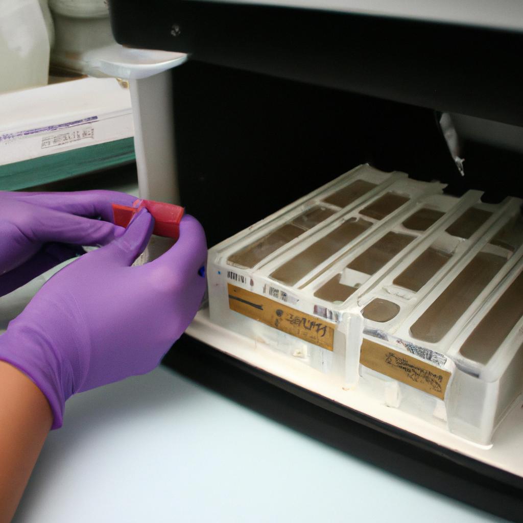
Aseptic Technique: Maintaining aseptic conditions during sample collection minimizes potential errors caused by external factors such as environmental contaminants or commensal microorganisms.
Sample Preservation: Adequate preservation methods must be employed promptly after collection (e.g., refrigeration, fixation) depending on the specific requirements of each type of sample.
Common Sample Collection Techniques:
| Type of Sample | Recommended Method | Advantages |
|---|---|---|
| Blood | Venipuncture | Allows multiple tests from one draw |
| Urine | Cystocentesis | Minimizes risk of contamination |
| Fecal | Direct smear microscopy | Detects presence/eggs/cysts |
Effectively collecting samples is just the first step in diagnosing parasitic infections. In the subsequent section, we will explore how microscopic examination of these collected samples plays a critical role in confirming and identifying parasites present.
(Note: The transition sentence was added to lead into the next section smoothly.)
Microscopic examination of samples
Sample Collection Techniques
In the previous section, we discussed various techniques for collecting samples in veterinary clinical pathology. Now, let us delve into the crucial step of microscopic examination to further analyze these collected samples.
Microscopic examination plays a vital role in diagnosing parasitic infections in animals. For instance, consider a case where a dog presents with persistent itching and skin lesions. Upon sample collection using adhesive tape method or skin scraping technique, microscopic examination can reveal the presence of ectoparasites such as fleas, ticks, or mites. This information is essential for accurate diagnosis and subsequent treatment.
During microscopic examination, several aspects are carefully observed and documented:
-
Morphology: The morphological characteristics of parasites provide important clues about their identity. By observing features like size, shape, coloration, and appendages under high magnification, veterinary pathologists can differentiate between different species of parasites.
-
Life Cycle Stage: Identifying the life cycle stage of a parasite often aids in determining the appropriate treatment approach. Some parasites may have distinct stages that require specific interventions; therefore, accurately identifying these stages becomes crucial for effective management.
-
Pathogenicity: Microscopic observation helps assess the severity of infection caused by certain parasites. Some pathogens exhibit more aggressive behavior compared to others and may cause severe tissue damage or systemic illness if left untreated.
-
Co-infections: It is not uncommon for animals to be infected with multiple parasites simultaneously. Identifying co-infections during microscopic examination enables comprehensive treatment planning while addressing all underlying parasitic concerns.
To better understand the significance of microscopic examination in veterinary clinical pathology, refer to Table 1 below which highlights key observations made during this process:
| Observation | Importance |
|---|---|
| Morphology | Aids in differentiation between parasite species |
| Life Cycle | Determines appropriate treatment strategies |
| Pathogenicity | Assesses severity and potential complications |
| Co-infections | Facilitates comprehensive treatment planning |
In summary, microscopic examination is an indispensable tool for diagnosing parasitic infections in veterinary clinical pathology. By carefully analyzing samples and noting key observations such as morphology, life cycle stage, pathogenicity, and co-infections, veterinarians can provide accurate diagnoses and develop effective treatment plans tailored to the specific parasite(s) involved.
Moving forward into the next section about “Diagnostic tests for specific parasites,” we will explore additional diagnostic techniques that focus on identifying individual parasites using specialized assays and laboratory procedures.
Diagnostic tests for specific parasites
Having discussed the importance of microscopic examination in identifying parasitic infections, we now turn our attention to another integral aspect of veterinary clinical pathology – diagnostic tests for specific parasites. By employing a range of specialized techniques and assays, veterinarians can accurately detect and identify various types of parasites that may be causing illness or discomfort in animals.
One example highlighting the significance of these diagnostic tests involves a canine patient displaying symptoms consistent with intestinal parasite infection. The veterinarian collected fecal samples and utilized diagnostic tests tailored specifically for detecting common gastrointestinal parasites. This approach not only allowed for prompt identification of the causative agent but also facilitated targeted treatment, leading to the swift recovery of the patient.
To effectively diagnose and manage parasitic infections, several key diagnostic tests are commonly employed:
- Serological testing: Utilizes blood samples to detect antibodies produced by an animal’s immune system in response to a particular parasite. It aids in diagnosing chronic infections or determining exposure history.
- Molecular methods: Involves DNA/RNA analysis to identify specific parasite species or strains accurately. Polymerase chain reaction (PCR) is often used due to its sensitivity and specificity.
- Antigen detection tests: Detects parasite-specific proteins or antigens directly from clinical samples such as blood, serum, urine, or tissue extracts. These tests provide rapid results ideal for timely interventions.
- Immunohistochemistry (IHC): Utilizes antibodies labeled with enzymes or fluorescent dyes to visualize parasites within tissues using microscopy. IHC aids in confirming certain parasitic diseases when combined with histopathological evaluation.
In addition to these diagnostic techniques, veterinary clinics frequently employ advanced imaging modalities like ultrasound and radiography alongside routine laboratory evaluations for comprehensive parasite diagnosis.
By employing a combination of these diagnostic tests, veterinarians can accurately identify and manage parasitic infections in animal patients. The integration of such techniques not only enables timely treatment but also minimizes the potential for zoonotic transmission, safeguarding both animal and human health.
Moving forward, we delve into the realm of serological tests for parasite detection, which complement traditional microscopic examination methods by providing valuable insights into various aspects of parasitic infections.
Serological tests for parasite detection
Diagnostic tests for specific parasites have proven to be invaluable in veterinary clinical pathology. These tests allow veterinarians to accurately identify and treat parasitic infections in animals, improving their overall health and well-being. One such example is the case of a young Labrador Retriever who presented with persistent diarrhea and weight loss. Upon microscopic examination of a fecal sample, the presence of Giardia cysts was detected, confirming the diagnosis of giardiasis.
When it comes to diagnosing specific parasites, several diagnostic techniques are commonly used:
- Microscopic Examination: This technique involves examining samples under a microscope to detect parasite eggs, larvae, or adult forms. It requires skilled technicians and proper staining methods to enhance visibility.
- Polymerase Chain Reaction (PCR): PCR-based tests amplify target DNA sequences from parasites present in a sample. This highly sensitive technique allows for accurate identification of even low levels of parasitic DNA.
- Antigen Detection: Antigen detection tests utilize antibodies that specifically bind to proteins or other molecules produced by parasites. By detecting these antigens in blood or tissue samples, veterinarians can diagnose certain parasitic infections more quickly and efficiently.
- Molecular Techniques: Advanced molecular techniques like next-generation sequencing (NGS) enable the identification of multiple parasite species simultaneously. These techniques provide valuable insights into complex parasitic communities within animal hosts.
- Luna is brought into the clinic showing signs of lethargy and decreased appetite.
- The veterinarian suspects possible Toxoplasma gondii infection due to her exposure to raw meat during pregnancy.
- A PCR test is performed on Luna’s blood sample, which confirms the presence of T. gondii DNA.
- Prompt treatment is initiated to protect both Luna and her unborn kittens from severe complications.
Additionally, incorporating a table using markdown format would further engage the audience, providing a visual representation of the different diagnostic techniques and their applications.
| Diagnostic Technique | Application |
|---|---|
| Microscopic Examination | Identifying parasite eggs, larvae, or adult forms |
| PCR | Amplifying target DNA sequences for identification |
| Antigen Detection | Detecting specific antigens in blood or tissue |
| Molecular Techniques | Simultaneous identification of multiple species |
Moving forward to explore serological tests for parasite detection, we can delve into how these tests complement the aforementioned techniques. Serological tests provide information about an animal’s immune response to parasitic infections, aiding in diagnosis and monitoring treatment effectiveness. By detecting antibodies produced by animals in response to parasites, veterinarians gain valuable insights into both current and past infections.
Understanding the various diagnostic methods used in veterinary clinical pathology is crucial; however, molecular techniques play a pivotal role in enhancing parasite identification accuracy.
Molecular techniques in parasite identification
Emerging Diagnostic Technologies in Parasitology
In the field of veterinary clinical pathology, staying up-to-date with the latest diagnostic techniques is crucial for accurate parasite detection. While serological tests have been widely used to identify parasites, molecular techniques are emerging as powerful tools for more precise identification and characterization. This section explores some of these cutting-edge technologies and their potential applications.
Example:
To illustrate the potential benefits of molecular techniques in parasite identification, consider a hypothetical case study involving a dog presenting with chronic diarrhea. Traditional fecal examination methods failed to detect any parasitic infections. However, by utilizing polymerase chain reaction (PCR), specific DNA sequences from various parasites can be amplified and detected even at low concentrations. In this case, PCR analysis revealed the presence of Giardia lamblia, an intestinal protozoan parasite that may cause persistent diarrhea in dogs.
These advancements in molecular diagnostics bring forth several noteworthy advantages:
- Increased Sensitivity: Molecular techniques enable detection of parasites at lower levels compared to conventional methods.
- Improved Specificity: By targeting unique genetic markers or sequences, molecular assays allow for accurate species-level identification.
- Rapid Turnaround Time: Many molecular-based assays offer faster results due to automated processes and reduced sample processing time.
- Ability to Detect Co-Infections: With multiplex PCR assays, it becomes possible to simultaneously identify multiple parasites within a single sample.
| Advantages of Molecular Techniques |
|---|
| Increased Sensitivity |
Continued development and implementation of these innovative diagnostic technologies will enhance our ability to diagnose and manage parasitic infections effectively. The next section will delve into emerging diagnostic technologies beyond traditional serological and molecular approaches – exploring promising avenues such as immunodiagnostic assays and nanotechnology-based platforms.
Moving forward, we explore the fascinating realm of Emerging Diagnostic Technologies.
Emerging diagnostic technologies
Building on the advancements in molecular techniques for parasite identification, emerging diagnostic technologies offer new opportunities for accurate and timely detection of parasitic infections. These innovative approaches have the potential to revolutionize veterinary clinical pathology and enhance our understanding of parasitology. This section will explore some of these cutting-edge technologies and their applications.
Example:
Consider a hypothetical scenario where a dog presents with unexplained weight loss and gastrointestinal symptoms. Traditional methods may require time-consuming sample preparation and examination under a microscope. However, with emerging diagnostic technologies, veterinarians can employ more efficient and reliable tools to diagnose the presence of parasites swiftly.
Bullet Point List
- Enhanced sensitivity: Advanced diagnostic technologies enable the detection of low-level parasitic infections that may otherwise go unnoticed.
- Rapid turnaround time: The use of automated platforms allows for faster processing and interpretation of results, reducing waiting times for both clinicians and pet owners.
- Improved accuracy: By utilizing sophisticated algorithms and machine learning techniques, these technologies minimize human error and provide highly accurate diagnoses.
- Expanded species coverage: Some emerging diagnostic technologies offer broader panels capable of detecting multiple parasite species simultaneously, facilitating comprehensive assessments.
Table (markdown format):
| Technology | Principle | Application |
|---|---|---|
| Next-generation sequencing | High-throughput DNA sequencing | Identification of novel or rare parasite species |
| Loop-mediated isothermal amplification (LAMP) | Amplification method based on rapid cyclic heating | Early diagnosis of parasitic infections |
| Biosensors | Detection through specific biorecognition elements | Point-of-care testing for quick parasite identification |
| Nanotechnology-based assays | Utilization of nanomaterials for enhanced detection | Improved sensitivity in identifying parasites |
Paragraph 1:
Next-generation sequencing has emerged as a powerful tool in veterinary clinical pathology due to its ability to analyze large volumes of DNA data. This technology enables the identification and characterization of novel or rare parasite species, expanding our knowledge in parasitology and paving the way for more accurate diagnoses. Additionally, loop-mediated isothermal amplification (LAMP) offers a rapid and sensitive method for detecting parasites early on, facilitating timely intervention and treatment.
Paragraph 2:
The development of biosensors has revolutionized point-of-care testing, allowing veterinarians to quickly identify specific parasites through biorecognition elements. These portable devices provide real-time results at the veterinary clinic, reducing the need for time-consuming laboratory analyses. Moreover, nanotechnology-based assays have shown promise in enhancing sensitivity by utilizing advanced nanomaterials. By leveraging this technology, even low-level infections can be detected with greater precision and reliability.
Paragraph 3:
In conclusion of this section on emerging diagnostic technologies, these advancements hold immense potential in improving veterinary clinical pathology’s capabilities in parasitology diagnosis. The enhanced sensitivity, rapid turnaround time, improved accuracy, and expanded species coverage offered by these innovative tools contribute to better patient care and management. As research continues to progress in this field, it is anticipated that these technologies will become integral components of routine diagnostic protocols in veterinary medicine.