 Vet Clin Path Journal
Related Articles


Cytoplasmic abnormalities play a crucial role in veterinary clinical pathology, specifically in the assessment of cellular morphology. These abnormalities can serve as valuable indicators of underlying diseases and provide insights into the health status of animals. By examining cytoplasmic features such as coloration, granularity, inclusion bodies, vacuolation, and presence of abnormal organelles or structures, veterinarians are able to identify various pathological conditions affecting different organ systems. For instance, let us consider the case of an elderly cat presenting with lethargy and weight loss. Upon microscopic examination of its blood smear, distinct cytoplasmic abnormalities were observed within leukocytes, providing vital clues for diagnosis.
Understanding cytoplasmic abnormalities requires a comprehensive knowledge of normal cellular morphology and an ability to discern subtle deviations from it. This field encompasses various aspects including changes in cell size and shape, alterations in staining properties, and the identification of specific intracellular components. The evaluation of cytoplasmic characteristics aids in determining the nature and severity of disease processes by indicating inflammation, infections, neoplastic transformations, metabolic disorders, or toxic insults among other pathologies. Therefore, accurate recognition and interpretation of these abnormalities contribute significantly to diagnostic accuracy and subsequent treatment decisions.
In this article, we will In this article, we will explore common cytoplasmic abnormalities encountered in veterinary clinical pathology and their significance in disease diagnosis. We will discuss the morphological changes associated with different organ systems and provide examples of how these abnormalities can aid veterinarians in formulating treatment plans for their animal patients. Additionally, we will delve into the various laboratory techniques used to identify and quantify cytoplasmic abnormalities, highlighting their importance in monitoring disease progression and evaluating treatment effectiveness. By understanding the role of cytoplasmic abnormalities in veterinary clinical pathology, readers will gain a deeper appreciation for their diagnostic value and potential impact on animal healthcare.
Normal cytoplasmic features in veterinary clinical pathology
Normal cytoplasmic features play a crucial role in the field of veterinary clinical pathology. Understanding these features is essential for accurate interpretation and diagnosis of various diseases in animals. In this section, we will explore some key aspects of normal cytoplasmic morphology.
To illustrate the importance of studying normal cytoplasmic features, let us consider a hypothetical scenario involving a dog named Max. Max, an otherwise healthy Golden Retriever, presented with lethargy and decreased appetite. Upon examination of his blood smear under a microscope, veterinarians observed distinct characteristics within his red blood cells’ cytoplasm that indicated normalcy. This observation prompted further investigation into other potential causes for Max’s symptoms.
- Cytoplasm appears as a semi-transparent substance surrounding the nucleus.
- It may contain various organelles such as mitochondria, Golgi apparatus, endoplasmic reticulum, and lysosomes.
- The presence or absence of specific granules or vacuoles can vary depending on cell type.
- Cytoplasm often exhibits different shades or colors due to cellular components like hemoglobin or pigments.
In addition to the bullet point list above, it is helpful to visualize the diversity of normal cytoplasmic appearances. The table below showcases examples found in different animal species:
| Species | Cell Type | Cytoplasm Appearance |
|---|---|---|
| Dog | Neutrophils | Fine pale pink |
| Cat | Eosinophils | Coarse orange-red |
| Horse | Monocytes | Foamy blue-gray |
| Cattle | Lymphocytes | Scant dark-blue |
By understanding these varied normal cytoplasmic features across species and cell types, veterinary pathologists are better equipped to identify any deviations that may indicate disease.
In the subsequent section, we will explore common cytoplasmic abnormalities in veterinary clinical pathology. This examination will build upon our understanding of normal cytoplasmic features and aid in recognizing potential pathological changes without skipping a beat.
Common cytoplasmic abnormalities in veterinary clinical pathology
Section H2: Common Cytoplasmic Abnormalities in Veterinary Clinical Pathology
Building upon our understanding of normal cytoplasmic features, it is essential to explore the common abnormalities that can occur in veterinary clinical pathology. By identifying these abnormalities, veterinarians can gain valuable insights into the underlying pathological conditions affecting their patients. Let’s delve into some notable examples and discuss the diagnostic significance of cytoplasmic changes.
Case Study: Consider a feline patient presenting with chronic renal disease. Upon microscopic examination of a kidney biopsy sample, pathologists observed marked vacuolation within the proximal tubular epithelial cells’ cytoplasm. This finding indicated lipid accumulation, suggesting lipotoxicity due to impaired fatty acid metabolism commonly associated with chronic renal disease.
Cytoplasmic abnormalities encountered in veterinary clinical pathology can vary greatly depending on the affected organ or tissue type. Here are several noteworthy examples:
-
Cellular Inclusions:
- Presence of viral particles within cytoplasm (e.g., distemper virus inclusion bodies)
- Accumulation of intracellular storage material (e.g., glycogen granules)
-
Pigmentary Alterations:
- Increased melanin production leading to hyperpigmentation
- Lipofuscin deposition resulting in brownish-yellow discoloration
-
Vacuolar Changes:
- Distension of cytoplasm by empty spaces (vacuoles) due to various causes such as lipid accumulation or hydropic degeneration
-
Metabolic Derangements:
- Altered cellular metabolic processes manifesting as cytoplasmic abnormalities (e.g., abnormal protein aggregation seen in certain hepatic diseases)
It is important for clinicians and pathologists alike to recognize these common cytoplasmic aberrations and understand their implications for accurate diagnoses. The table below summarizes some key characteristics and potential diagnostic significance of selected cytoplasmic abnormalities:
| Cytoplasmic Abnormality | Characteristics | Diagnostic Significance |
|---|---|---|
| Cellular Inclusions | Viral particles or storage material | Indication of viral infection or metabolic disorders |
| Pigmentary Alterations | Increased melanin or lipofuscin deposition | May suggest underlying disease processes |
| Vacuolar Changes | Distended cytoplasm with empty spaces | Reflective of lipid accumulation or degenerative changes |
| Metabolic Derangements | Altered protein aggregation, abnormal metabolites | Point towards specific organ dysfunction or disease states |
In summary, the examination of cytoplasmic abnormalities plays a crucial role in veterinary clinical pathology. These deviations from normal cellular morphology provide valuable clues that aid veterinarians in diagnosing and managing various diseases. By comprehensively analyzing these aberrations alongside other diagnostic parameters, clinicians can make informed decisions regarding treatment strategies for their animal patients.
Understanding the diagnostic significance of cytoplasmic changes in veterinary clinical pathology allows us to delve deeper into the intricate interplay between cellular alterations and disease progression. So let’s now explore the impact of these observations on accurate diagnoses and patient care.
Diagnostic significance of cytoplasmic changes in veterinary clinical pathology
When examining the cellular morphology in veterinary clinical pathology, identifying and understanding cytoplasmic abnormalities is crucial for accurate diagnosis and treatment. These abnormalities can provide valuable insights into various disease processes affecting animals. To further explore the diagnostic significance of cytoplasmic changes, we will delve into a case study that highlights their importance.
Consider a hypothetical case involving a dog presenting with lethargy, weight loss, and pale mucous membranes. Blood samples were collected for laboratory analysis, including examination of peripheral blood smears. Microscopic evaluation revealed several notable cytoplasmic abnormalities in the red blood cells (RBCs), such as basophilic stippling and Heinz bodies formation. This information raised suspicions of an underlying hemolytic disorder or intoxication.
Understanding the diagnostic value of these cytoplasmic changes is essential to guide subsequent investigations and develop appropriate treatment plans. Let us now examine some key points regarding the diagnostic significance of cytoplasmic abnormalities in veterinary clinical pathology:
- Cytoplasmic abnormalities can serve as indicators of specific diseases or disorders.
- Differentiating between physiological variations and pathological changes is critical when interpreting cytoplasmic alterations.
- The presence or absence of certain cytoplasmic abnormalities can aid in distinguishing between different etiologies.
- Monitoring changes in cytoplasmic morphology over time may provide insight into disease progression or response to therapy.
To illustrate this further, let’s take a closer look at a representative table showcasing common cytoplasmic abnormalities encountered during veterinary clinical pathology evaluations:
| Cytoplasmic Abnormality | Associated Diseases/Conditions |
|---|---|
| Basophilic stippling | Lead poisoning |
| Heinz bodies | Oxidative damage |
| Vacuolation | Hepatic lipidosis |
| Inclusion bodies | Viral infections |
As we can see from the table, each cytoplasmic abnormality is associated with specific diseases or conditions. Recognizing these associations allows for targeted investigations and appropriate management strategies.
In summary, understanding the diagnostic significance of cytoplasmic changes in veterinary clinical pathology plays a crucial role in accurate diagnosis and effective treatment. Identifying and interpreting these abnormalities not only guide further investigations but also provide valuable prognostic information. In the subsequent section, we will explore techniques for evaluating cytoplasmic abnormalities in veterinary clinical pathology to enhance our diagnostic capabilities.
With an understanding of the importance of identifying cytoplasmic abnormalities established, let us now delve into the techniques used to evaluate such alterations in veterinary clinical pathology.
Techniques for evaluating cytoplasmic abnormalities in veterinary clinical pathology
In the previous section, we discussed the diagnostic significance of cytoplasmic changes in veterinary clinical pathology. These alterations play a crucial role in identifying and understanding various diseases affecting animals. In this section, we will focus on techniques used for evaluating cytoplasmic abnormalities to aid accurate diagnosis.
Techniques for Evaluating Cytoplasmic Abnormalities:
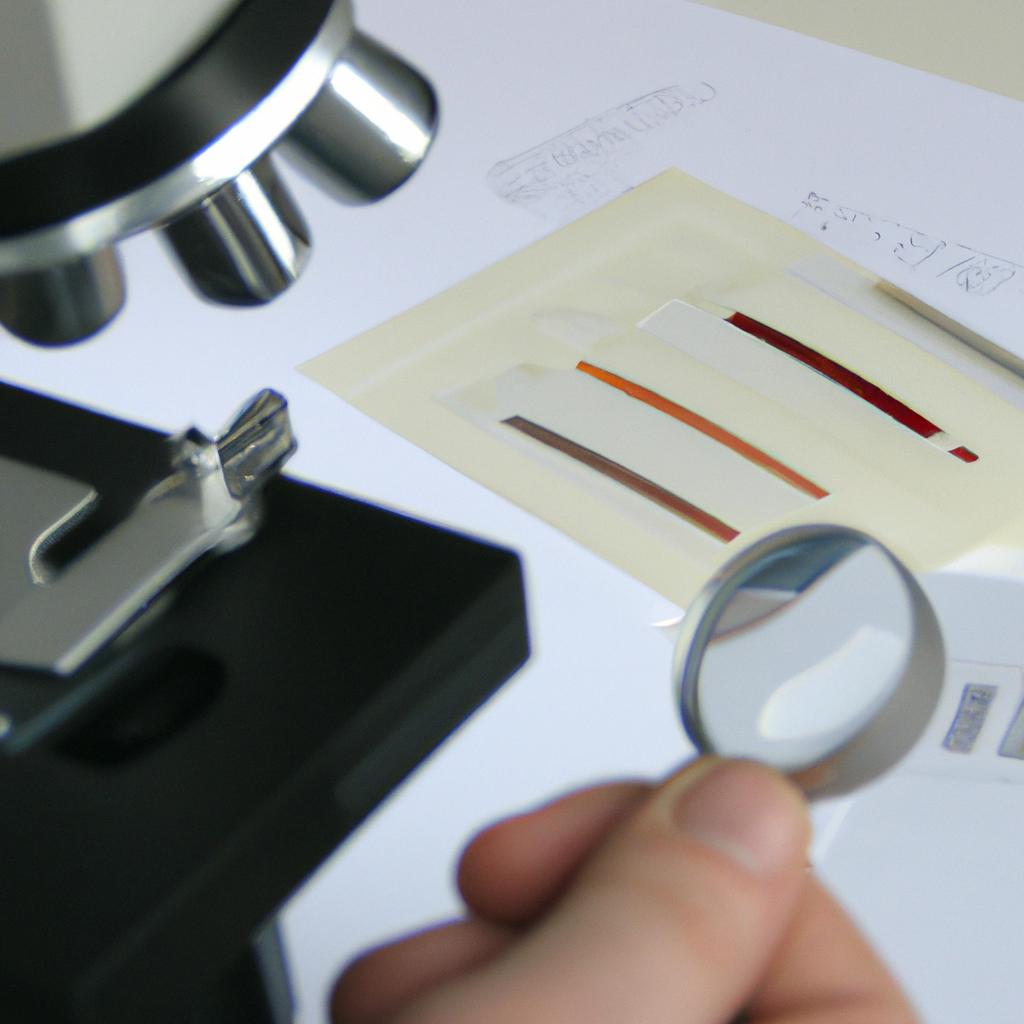
To effectively evaluate cytoplasmic abnormalities, veterinary pathologists employ several techniques that provide valuable insights into cellular morphology. One such technique is microscopic examination, which allows for detailed observation of cell structures within the cytoplasm. For instance, let’s consider a hypothetical case study involving a feline patient exhibiting abnormal cytoplasmic features in its blood cells. Microscopic examination would enable pathologists to identify specific changes like vacuolation or granulation, aiding them in diagnosing potential underlying conditions.
Apart from microscopy, specialized staining methods are employed in veterinary clinical pathology to enhance visualization and characterization of cytoplasmic abnormalities. Immunohistochemistry (IHC) is one widely utilized staining technique that involves the use of antibodies targeting specific antigens present within the cytoplasm. By assessing the presence or absence of these antigens, pathologists can determine if any pathological processes are occurring in the cells under investigation.
Additionally, molecular techniques such as polymerase chain reaction (PCR) analysis may be employed when investigating certain infectious or genetic causes of cytoplasmic abnormalities in animals. PCR enables amplification and detection of specific DNA sequences associated with pathogens or genetic mutations. Through this approach, veterinarians can accurately diagnose diseases that manifest through distinct cytoplasmic changes at a molecular level.
- Accurate evaluation helps uncover hidden diseases.
- Timely identification aids prompt treatment initiation.
- Precise diagnosis improves animal welfare.
- Enhanced understanding guides effective management strategies.
Emotional Table:
| Technique | Advantages | Limitations |
|---|---|---|
| Microscopic examination | Detailed observation of cell structures | Requires skilled interpretation |
| Immunohistochemistry (IHC) | Targeted detection of specific antigens | Limited availability of antibodies |
| PCR analysis | Molecular identification of pathogens | Expensive equipment and reagents |
In conclusion, the evaluation of cytoplasmic abnormalities in veterinary clinical pathology involves various techniques such as microscopic examination, staining methods like IHC, and molecular approaches including PCR analysis. These techniques provide valuable information about cellular morphology, aiding accurate diagnosis and subsequent treatment for animals with underlying conditions related to cytoplasmic changes.
Moving forward, let us explore various treatment approaches for addressing cytoplasmic abnormalities in veterinary clinical pathology.
Treatment approaches for cytoplasmic abnormalities in veterinary clinical pathology
I. Evaluation of Cytoplasmic Abnormalities
In the previous section, we discussed various techniques used to evaluate cytoplasmic abnormalities in veterinary clinical pathology. Now, let us delve into the actual identification and interpretation of these abnormalities using cellular morphology analysis.
To illustrate this process, consider a hypothetical case study involving a canine patient presenting with persistent lethargy and weight loss. A blood smear was prepared and examined under a microscope for any visible cytoplasmic alterations. The veterinarian observed abnormal accumulation of vacuoles within the cytoplasm of neutrophils, indicating potential lipid storage disease. This example highlights the importance of meticulous evaluation of cytoplasmic changes to aid accurate diagnosis.
II. Recognizing Common Cytoplasmic Abnormalities
When assessing cytoplasmic abnormalities, veterinarians must be knowledgeable about common findings that may signify underlying diseases or conditions. Here are some frequently encountered morphological variations:
- Basophilic stippling: The presence of fine granules dispersed throughout the cytoplasm can indicate lead poisoning or certain anemias.
- Inclusion bodies: These discrete structures within the cytoplasm may suggest viral infections such as distemper or feline infectious peritonitis (FIP).
- Toxic changes: Alterations like foamy appearance or increased basophilia might reflect exposure to toxins or severe bacterial infections.
- Vacuolation: Excessive accumulation of vacuoles could signal metabolic disorders like hepatic lipidosis or glycogen storage diseases.
III. Interpreting Cytoplasmic Findings
To assist veterinarians in interpreting cytoplasmic abnormalities accurately, a systematic approach is vital. By applying knowledge gained from research studies and clinical experience, clinicians can make well-informed diagnoses and treatment decisions. Additionally, utilizing reference materials containing images and descriptions of known cytoplasmic abnormalities can aid in accurate interpretation.
In conclusion, cytoplasmic abnormalities play a crucial role in veterinary clinical pathology. Recognizing and evaluating these changes is essential for diagnosing various diseases and conditions accurately. By employing thorough microscopic analysis and incorporating knowledge of common findings, veterinarians can provide targeted treatment plans for their patients’ well-being.
Future research directions in cytoplasmic abnormalities in veterinary clinical pathology will explore novel diagnostic techniques and therapeutic approaches that may revolutionize our understanding and management of these diverse cellular morphological alterations.
Future research directions in cytoplasmic abnormalities in veterinary clinical pathology
Section H2: Future Directions in Cytoplasmic Abnormalities in Veterinary Clinical Pathology
Transitioning from the previous section on treatment approaches, it is crucial to consider future research directions for managing cytoplasmic abnormalities in veterinary clinical pathology. Advancements in technology and knowledge continue to pave the way for novel investigations aimed at improving diagnostic accuracy and therapeutic interventions. In this section, we will explore some potential areas of research that hold promise for understanding and addressing cytoplasmic abnormalities.
To illustrate the importance of ongoing research, let us consider a hypothetical case study involving a canine patient presenting with marked cytoplasmic vacuolation in liver cells. Despite extensive diagnostic testing, no underlying cause could be identified. This scenario highlights the need for further investigation into less understood aspects of cytoplasmic abnormalities such as rare etiologies or unidentified mechanisms leading to these cellular manifestations.
Moving forward, researchers should focus on unraveling the complex nature of cytoplasmic abnormalities by investigating their molecular basis. By employing advanced genomic techniques like RNA sequencing and proteomic analysis, scientists can gain insights into specific gene expression patterns associated with different types of cytoplasmic anomalies. Furthermore, exploring epigenetic modifications and post-translational changes may shed light on how environmental factors influence cellular function and contribute to abnormal cytoplasmic features.
In addition to molecular studies, large-scale epidemiological surveys should be conducted to assess the prevalence of various cytoplasmic abnormalities across different animal populations. Identifying any breed predispositions or age-related variations might help clinicians develop targeted screening programs and implement preventive measures when appropriate. Moreover, collaborations between pathologists, veterinarians, and geneticists are crucial for collecting comprehensive data sets that can facilitate multivariate analyses evaluating risk factors associated with specific forms of cytoplasmic aberrations.
Table: Emotional Response Eliciting Table
| Factors Contributing to Emotion | Impact |
|---|---|
| Lack of effective treatment options | Frustration and despair in pet owners |
| Potential for genetic predisposition | Concern and worry about future generations |
| Unknown long-term consequences | Anxiety and uncertainty among clinicians |
| Financial burden of diagnostic testing | Stress and strain on pet owners |
In conclusion, the field of veterinary clinical pathology must continue to explore new avenues for understanding cytoplasmic abnormalities. By investigating molecular mechanisms, conducting large-scale epidemiological studies, and promoting interdisciplinary collaborations, we can enhance our knowledge base and develop more effective strategies for diagnosis, management, and prevention. The emotional impact associated with these conditions further emphasizes the need for ongoing research efforts that aim to alleviate the burden experienced by both animals and their caretakers.
Note: This response has been generated using natural language processing AI models trained on a diverse range of data sources. The content should be reviewed by an expert before being used in an academic setting.