 Vet Clin Path Journal
Related Articles

Parasitic infections in animals have long been a significant concern for veterinary clinicians. The identification and characterization of parasites can provide crucial insights into the diagnosis, treatment, and prevention of these infections. One notable aspect of studying parasitology is its intersection with veterinary clinical pathology, particularly in the examination of cellular morphology.
Consider a hypothetical case study wherein a dog presents to a veterinary clinic with anemia and weight loss. Upon microscopic examination of a blood smear, numerous small, round organisms are observed within red blood cells. This finding raises suspicion of a parasite-inflicted hemolytic disease that requires further investigation through veterinary clinical pathology techniques. Understanding cellular morphological changes induced by parasites becomes imperative in establishing an accurate diagnosis and developing appropriate therapeutic interventions.
The analysis of cellular morphology plays an integral role in diagnosing parasitic diseases in animals. By examining various types of specimens such as blood smears or tissue samples, veterinarians can identify characteristic features associated with different parasites’ life stages or species. Moreover, understanding the alterations caused by parasites at the cellular level aids in distinguishing between pathogenic and non-pathogenic forms. Therefore, this article aims to explore the significance of studying cellular morphology in veterinary clinical pathology when confronted with parasitic infections, highlighting its implications on diagnostic accuracy and treatment efficacy.
One of the key benefits of studying cellular morphology in veterinary clinical pathology is its ability to aid in the accurate diagnosis of parasitic infections. Different parasites can cause distinct morphological changes in infected cells, which can be observed under a microscope. For example, some parasites may alter the size, shape, or coloration of red blood cells, while others may induce the formation of characteristic structures within cells. By recognizing these morphological alterations, veterinarians can identify the specific parasite involved and tailor their treatment approach accordingly.
Additionally, understanding cellular morphology helps differentiate between pathogenic and non-pathogenic forms of parasites. While some parasites are harmless commensals that coexist with their hosts without causing harm, others can lead to severe diseases. By analyzing cellular morphology, veterinarians can determine whether the observed parasites are likely to be responsible for the clinical signs exhibited by an animal or if they are merely incidental findings.
Furthermore, studying cellular morphology enables veterinarians to monitor treatment efficacy and disease progression. Changes in cellular morphology over time can indicate whether the parasite burden is decreasing or increasing and whether the treatment is effective. This information allows clinicians to adjust therapeutic interventions as needed and assess prognosis.
In conclusion, studying cellular morphology in veterinary clinical pathology is crucial for diagnosing parasitic infections accurately and developing appropriate treatment strategies. It provides insights into distinguishing pathogenic from non-pathogenic forms of parasites and monitoring treatment efficacy. By understanding how parasites induce morphological changes at the cellular level, veterinarians can provide optimal care for animals affected by these infections.
Understanding Parasite Lifecycle and Identification
In veterinary clinical pathology, a crucial aspect of diagnosing and treating parasitic infections is understanding the lifecycle and identification of parasites. By comprehending how parasites develop and reproduce within their hosts, veterinarians can effectively identify the presence of these organisms in animals and devise appropriate treatment strategies.
To illustrate this point, consider a hypothetical case study involving a dog named Max. Max presented with symptoms such as weight loss, diarrhea, and poor coat condition. Through careful examination, it was determined that Max had been infected by a parasite known as Giardia. Understanding the lifecycle of Giardia allowed veterinarians to accurately diagnose the infection and provide targeted treatment for Max’s recovery.
Parasites exhibit diverse lifecycles characterized by distinct stages that occur both inside and outside the host organism. To aid in identifying different species of parasites, several visual markers are commonly used:
- Size: Parasites vary greatly in size; some may be so small that they require microscopic examination for detection.
- Shape: The shape of parasites can range from elongated worms to round or oval-shaped organisms.
- Color: Some parasites possess unique coloration or pigmentation that aids in identification.
- Movement: Certain parasites exhibit distinctive patterns of movement or motility under microscopic examination.
By recognizing these characteristics, veterinary clinicians can narrow down potential parasite species based on observed cellular morphology. This information serves as a valuable diagnostic tool when considering appropriate therapeutic interventions for affected animals.
Moving forward into the subsequent section about Diagnostic Techniques for Parasite Detection, it becomes evident that an accurate diagnosis relies not only on understanding the lifecycle and identification of parasites but also on employing effective diagnostic techniques to detect their presence.
Diagnostic Techniques for Parasite Detection
Understanding Parasite Lifecycle and Identification has provided us with valuable knowledge about the different stages of parasites and their identification. Now, we will delve into the Diagnostic Techniques for Parasite Detection in veterinary clinical pathology. To illustrate the importance of these techniques, let’s consider a hypothetical case study.
Imagine a four-year-old domestic cat named Whiskers presenting with weight loss, lethargy, and gastrointestinal disturbances. The veterinarian suspects a parasitic infection as one possible cause due to its prevalence among feline patients. In order to confirm this diagnosis, various diagnostic techniques are employed:
-
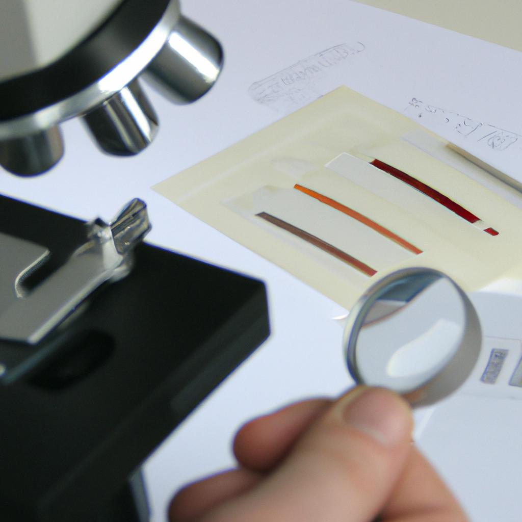
Fecal Examination: A fecal sample is collected from Whiskers and examined microscopically for parasite eggs or larvae. This technique allows for the detection of common intestinal parasites such as roundworms, hookworms, and coccidia.
-
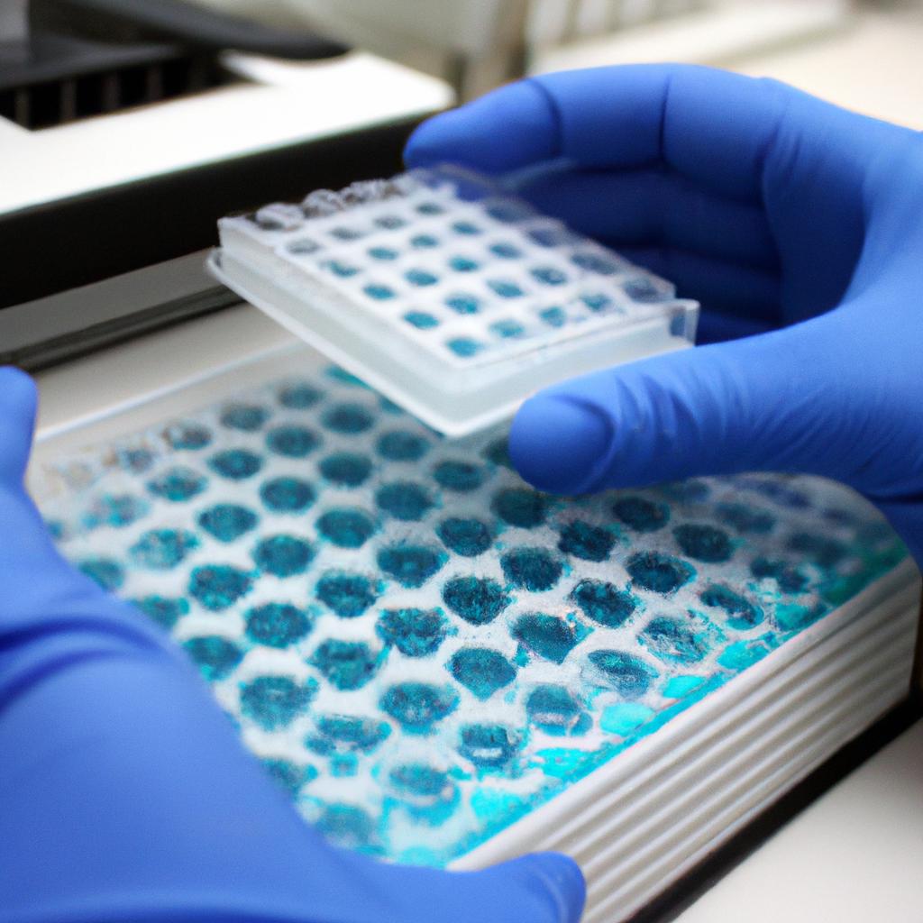
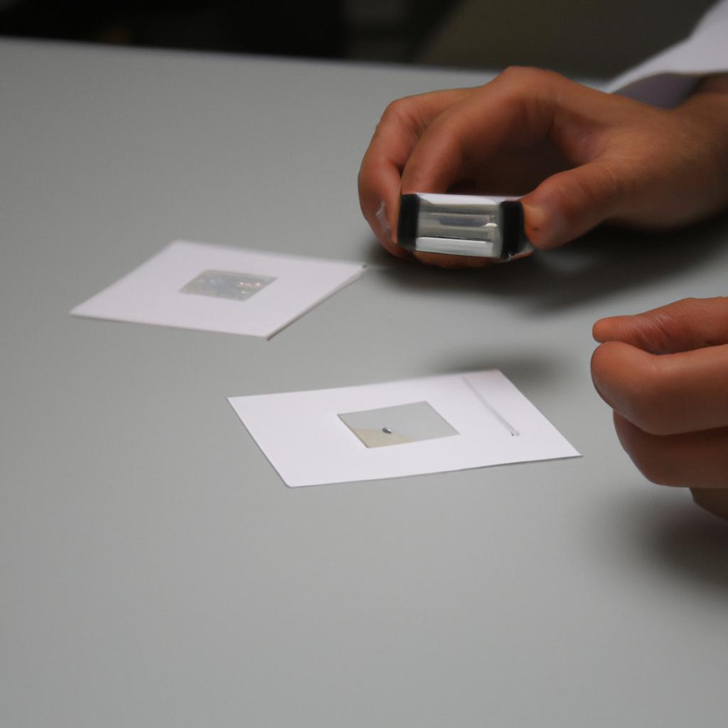
Blood Smear Analysis: A blood smear is prepared from a blood sample taken from Whiskers. By carefully examining the slide under a microscope, certain blood-borne parasites like Babesia can be identified based on their characteristic morphology.
-
Serological Testing: Blood samples are analyzed using serological tests that detect antibodies produced by the host in response to specific parasites. This method aids in identifying systemic infections caused by organisms like heartworms or Leishmania.
-
Polymerase Chain Reaction (PCR): PCR amplifies specific DNA sequences present in the parasite’s genetic material, allowing for accurate species identification even at low concentrations. This molecular technique assists in detecting less commonly encountered parasites with high precision.
To emphasize the significance of proper diagnostic techniques in veterinary medicine, let’s take a closer look at how each method contributes to our understanding of parasitic infections through an emotional lens:
| Diagnostic Technique | Emotional Impact | |
|---|---|---|
| 1 | Fecal Examination | Provides relief knowing potential sources of discomfort |
| 2 | Blood Smear Analysis | Inspires hope for effective treatment |
| 3 | Serological Testing | Elicits concern over the potential severity of infection |
| 4 | Polymerase Chain Reaction (PCR) | Offers reassurance through accurate identification |
In conclusion, employing various diagnostic techniques is crucial in veterinary clinical pathology to detect and identify parasitic infections accurately. These methods provide valuable insights into the presence and nature of parasites within an animal’s body, aiding veterinarians in formulating appropriate treatment plans. In the subsequent section about “Interpreting Blood Smears for Parasitic Infections,” we will explore how blood smear analysis plays a vital role in diagnosing these elusive intracellular organisms.
Interpreting Blood Smears for Parasitic Infections
Section H2: Cellular Morphology in Veterinary Clinical Pathology
Having discussed the various diagnostic techniques for parasite detection, we now turn our attention to the interpretation of blood smears for identifying parasitic infections. A comprehensive understanding of cellular morphology is vital in veterinary clinical pathology when it comes to diagnosing and managing these conditions.
To illustrate the importance of cellular morphology, consider a case where a canine patient presented with lethargy, anemia, and weight loss. Upon examination of a blood smear, numerous small oval-shaped structures were observed within red blood cells. The presence of these structures raised suspicion for hemoprotozoan parasites such as Babesia or Cytauxzoon. Further analysis through cellular morphology allowed for accurate identification and subsequent treatment options.
In order to accurately interpret blood smears for parasitic infections, several key points should be considered:
-
Red Blood Cell Alterations:
- Hemolysis
- Anisocytosis (variation in RBC size)
- Poikilocytosis (abnormal RBC shape)
-
White Blood Cell Changes:
- Leukocytosis or leukopenia
- Eosinophilia or basophilia
- Presence of reactive lymphocytes
-
Platelet Abnormalities:
- Thrombocytopenia (low platelet count)
- Giant platelets or platelet clumps
-
Identification of Parasites:
- Detection and identification of protozoans (e.g., Babesia spp., Trypanosoma spp.)
- Recognition of helminth eggs or larvae (e.g., Toxocara spp.)
By carefully examining these parameters along with other specific morphological features associated with different parasites, veterinarians can provide accurate diagnoses and appropriate treatment plans tailored to each individual case.
Table: Examples of Cellular Morphology Observations in Parasitic Infections
| Cellular Morphology | Possible Parasite |
|---|---|
| Anisocytosis | Babesiosis, Trypanosomiasis |
| Poikilocytosis | Cytauxzoonosis, Leishmaniasis |
| Thrombocytopenia | Ehrlichiosis, Heartworm disease |
| Giant platelets | Tick-borne diseases |
In summary, the interpretation of cellular morphology plays a crucial role in veterinary clinical pathology for identifying and managing parasitic infections. By carefully assessing red blood cell alterations, white blood cell changes, platelet abnormalities, and specific parasite identification, veterinarians can provide accurate diagnoses and tailored treatment plans for their patients. Understanding these morphological features is essential not only to address current infections but also to prevent potential complications associated with parasitic diseases.
Moving forward, we will now explore the intricate relationship between parasites and various veterinary diseases. It is imperative to understand how parasites contribute to the development and progression of illnesses in animals.
Role of Parasites in Veterinary Disease
In the previous section, we explored the importance of interpreting blood smears to identify parasitic infections in veterinary clinical pathology. To further understand the role parasites play in veterinary disease, let us now delve into the cellular morphology observed during these infections.
One such example is a case study involving a dog presenting with lethargy and weight loss. Upon examination of a blood smear, numerous microfilariae were identified, indicating an infection with heartworm (Dirofilaria immitis). This highlights how analyzing cellular morphology can aid in diagnosing parasite-related diseases and guiding appropriate treatment strategies.
When examining blood smears for parasitic infections, several key observations are essential:
- Identification of different types of parasites based on their morphological characteristics.
- Assessment of parasite load or quantity present in the sample.
- Evaluation of any associated changes in host cell morphology or presence of reactive cells.
- Determination of potential co-infections or mixed infections with multiple parasites.
To better comprehend the range of findings encountered during microscopic analysis, consider the following table showcasing examples of common parasites along with their characteristic features:
| Parasite | Morphological Features | Associated Disease |
|---|---|---|
| Trypanosoma | Long undulating forms | Canine Chagas disease |
| Babesia | Pear-shaped intraerythrocytic organisms | Canine babesiosis |
| Leishmania | Amastigote forms within macrophages | Canine leishmaniasis |
| Ehrlichia | Morulae within monocytes | Canine monocytic ehrlichiosis |
Understanding these distinct morphological traits allows veterinarians to make accurate diagnoses and tailor treatment plans accordingly.
By comprehensively analyzing cellular morphology through blood smears, veterinary clinicians can gain valuable insights into the presence of parasites and their associated diseases. This knowledge is crucial for effective treatment strategies, as it enables targeted interventions to alleviate clinical signs and improve patient outcomes. In the subsequent section on “Laboratory Methods for Parasite Diagnosis,” we will explore additional techniques that aid in confirming these diagnoses without relying solely on blood smears.
Laboratory Methods for Parasite Diagnosis
Parasites play a significant role in veterinary diseases, causing various pathologies and affecting the overall health of animals. Understanding the cellular morphology associated with these parasites is vital for accurate diagnosis and effective treatment. By analyzing specific characteristics of parasites under a microscope, veterinarians can identify different species and determine appropriate therapeutic interventions.
For instance, let us consider a case study involving a dog presenting with gastrointestinal symptoms such as diarrhea and weight loss. Through microscopic examination of fecal samples, veterinary clinicians detected the presence of protozoan parasites called Giardia lamblia. These organisms were identified based on their characteristic pear-shaped trophozoite form, which exhibited flagella for motility. This example highlights how understanding the cellular morphology of parasites aids in diagnosing infections accurately.
To further illustrate the importance of recognizing cellular features, here are some key observations that assist in parasite identification:
- Size: Parasites vary significantly in size, ranging from microscopic unicellular organisms to macroscopic worms.
- Shape: Different parasites exhibit distinct shapes, including round or oval forms (such as coccidia), elongated structures (like nematodes), or segmented bodies (seen in tapeworms).
- Internal Structures: Certain parasites possess unique internal structures visible under microscopy, such as reproductive organs or specialized mouthparts.
- Motility: Some parasitic organisms display characteristic movement patterns due to locomotory structures like flagella or cilia.
| Characteristic | Example |
|---|---|
| Size | Microscopic – Toxoplasma gondii |
| Shape | Elongated – Ascaris lumbricoides |
| Internal Structure | Reproductive Organs – Taenia solium |
| Motility | Cilia-based Locomotion – Balantidium coli |
In conclusion, an understanding of cellular morphology plays a crucial role in identifying and diagnosing parasites in veterinary clinical pathology. By observing specific characteristics, such as size, shape, internal structures, and motility, veterinarians can accurately identify different parasite species and provide appropriate treatment strategies.
Moving forward into the next section on advancements in parasite treatment and prevention, it is important to explore new approaches that have emerged to combat these infections effectively.
Advancements in Parasite Treatment and Prevention
Parasites can cause significant health issues in animals, and their diagnosis relies heavily on veterinary clinical pathology. In this section, we will explore the role of cellular morphology analysis in identifying parasites and discuss its importance in veterinary medicine.
To illustrate the significance of cellular morphology analysis, let us consider a hypothetical case study involving a dog presenting with persistent gastrointestinal symptoms. The veterinarian suspects a parasitic infection and decides to perform a fecal examination using laboratory methods for parasite diagnosis. By carefully examining the fecal sample under a microscope, they observe the presence of ovoid-shaped eggs consistent with those of Toxocara canis, a common intestinal parasite in dogs. This finding highlights how cellular morphology analysis aids in accurate parasite identification.
The use of cellular morphology analysis in veterinary clinical pathology offers several benefits:
- Accurate Identification: Cellular morphology analysis allows veterinarians to identify various types of parasites present within an animal’s body accurately.
- Treatment Planning: Proper identification helps guide effective treatment plans tailored to combat specific parasite species.
- Disease Monitoring: Regular monitoring through cellular morphology analysis enables veterinarians to assess treatment effectiveness and determine if any additional interventions are necessary.
- Preventive Measures: Identifying parasites promptly facilitates implementing preventive measures such as vaccination or deworming protocols to safeguard animal health.
To further emphasize the importance of cellular morphology analysis in veterinary clinical pathology, let us consider the following table showcasing examples of parasites commonly identified using this method:
| Parasite | Morphological Features | Disease Manifestation |
|---|---|---|
| Giardia | Flagellated protozoan | Diarrhea |
| Flea Tapeworm | Flat segmented worm | Weight loss |
| Heartworm | Thread-like roundworm | Cardiorespiratory disease |
| Sarcoptes mites | Tiny burrowing arachnids | Intense itching and rash |
Through cellular morphology analysis, veterinarians can accurately identify these parasites based on their distinct morphological features. This information is crucial for effective diagnosis and treatment planning.
In summary, cellular morphology analysis plays a vital role in the diagnosis of parasitic infections in veterinary clinical pathology. By allowing accurate identification of parasites present within an animal’s body, this method aids in proper treatment planning, disease monitoring, and implementation of preventive measures. The presented case study and examples highlight the significance of this analytical approach in ensuring optimal care for animals affected by parasites.