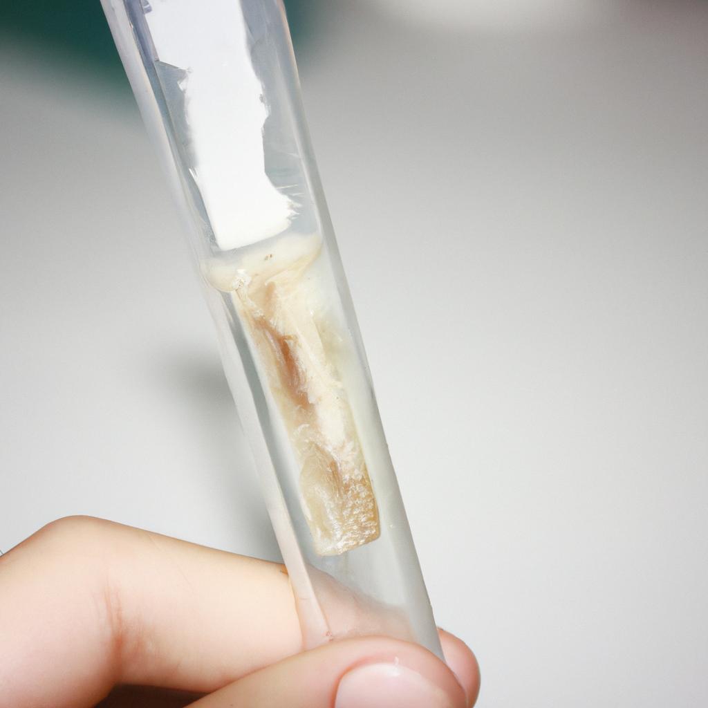
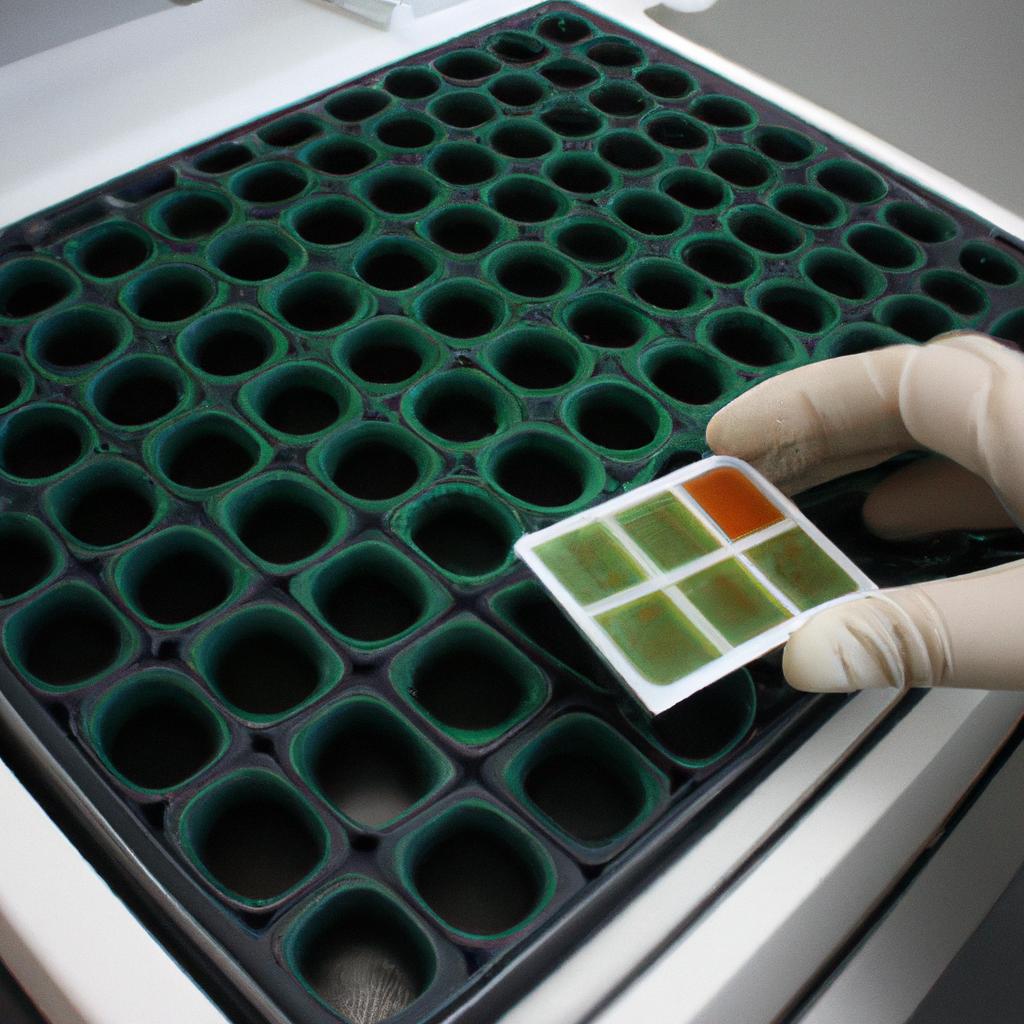
Veterinary clinical pathology is a crucial aspect of veterinary medicine, providing valuable insights into the health and well-being of animals. One area within this field that warrants special attention is hematology, which involves the study of blood cells and their components. By analyzing various parameters such as red blood cell count, White Blood Cell Differential, and platelet counts, veterinary clinicians can gain important diagnostic information to aid in disease detection and monitoring.
For instance, consider the case of a 10-year-old domestic shorthair cat presenting with lethargy and decreased appetite. Through a comprehensive hematological analysis, it was revealed that the cat had severe anemia characterized by significantly reduced red blood cell count and hemoglobin concentration. Further examination indicated the presence of macrocytic hypochromic erythrocytes, suggesting underlying nutritional deficiencies or chronic diseases affecting erythropoiesis. This example demonstrates how hematology provides critical clues about an animal’s overall health status while helping veterinarians narrow down potential causes for observed abnormalities.
Hematology serves as not only a diagnostic tool but also plays a pivotal role in monitoring response to treatment and assessing prognosis. In this article, we will delve deeper into the world of veterinary clinical pathology: hematology insights. We will explore various hemat ology parameters, their significance, and how they can be interpreted in different clinical scenarios. Additionally, we will discuss the importance of hematology in guiding treatment decisions and evaluating the effectiveness of interventions.
One important aspect of hematology is the evaluation of red blood cell parameters. These include red blood cell count (RBC), hemoglobin concentration (Hb), hematocrit (HCT), mean corpuscular volume (MCV), mean corpuscular hemoglobin (MCH), and mean corpuscular hemoglobin concentration (MCHC). Abnormalities in these parameters can provide insights into various conditions such as anemia, polycythemia, and certain underlying diseases.
White blood cell differential is another crucial component of a complete blood count analysis. It involves identifying and quantifying different types of white blood cells, including neutrophils, lymphocytes, monocytes, eosinophils, and basophils. An imbalance or abnormal increase/decrease in any particular cell type can indicate inflammation, infection, immune system disorders, or even certain types of cancer.
Platelet counts are also essential for assessing clotting function and monitoring potential bleeding disorders. Low platelet counts (thrombocytopenia) may result in prolonged bleeding or impaired clot formation, while high platelet counts (thrombocytosis) may indicate underlying inflammatory conditions or bone marrow disorders.
Besides these primary parameters, veterinary clinicians often evaluate additional indices such as reticulocyte count to assess bone marrow response to anemia and erythrocyte sedimentation rate (ESR) to measure the presence of inflammation.
Interpreting these hematological findings requires knowledge of normal reference ranges for different species and consideration of factors such as age, breed predispositions, concurrent medications/therapies, and patient history. It is important to note that hematological abnormalities alone do not provide a definitive diagnosis but rather serve as valuable screening tools that guide further investigations and diagnostic tests.
In conclusion, hematology is a vital component of veterinary clinical pathology that enables veterinarians to gain valuable insights into an animal’s health status. By analyzing various hematological parameters, clinicians can identify potential diseases or abnormalities, monitor treatment response, and evaluate prognosis. Understanding the significance of these parameters and their interpretation in different clinical scenarios allows for more accurate diagnosis and targeted interventions to improve animal welfare.
Importance of Hematology in Veterinary Medicine
Hematology, the study of blood and its components, plays a crucial role in veterinary medicine. By analyzing various parameters within the blood, veterinarians gain valuable insights into an animal’s overall health status and can diagnose a wide range of conditions. For instance, consider the case of a dog presenting with lethargy and loss of appetite. Through a comprehensive hematology evaluation, including a complete blood count (CBC) and examination of peripheral blood smears, veterinarians may identify anemia or abnormal white blood cell counts as potential underlying causes.
Understanding the importance of hematology requires recognition of its multifaceted applications. Firstly, it aids in disease detection by identifying deviations from normal ranges for red blood cells (RBCs), white blood cells (WBCs), platelets, and other cellular elements. These variations can indicate diseases such as infections, immune-mediated disorders, or cancers. Secondly, hematology provides essential information about an animal’s response to therapy by monitoring changes in cell populations over time. This allows veterinarians to assess treatment effectiveness and make necessary adjustments if required.
To emphasize the significance of hematology further, let us briefly explore some key benefits:
- Early Disease Detection: Hematological analysis enables early identification of diseases before clinical signs become apparent.
- Monitoring Treatment Progress: Regular assessment of hematologic parameters helps track an animal’s response to therapy and adjust treatments accordingly.
- Predictive Prognostication: Certain hematologic abnormalities provide prognostic indicators that aid in determining the likely outcome for specific conditions.
- Research Advancements: Hematological studies contribute significantly to scientific research on animal health and allow for advancements in diagnostic techniques and therapeutic interventions.
In addition to these advantages, understanding blood cell composition is fundamental to comprehending how different diseases affect animals differently based on their species-specific characteristics. The subsequent section will delve deeper into this topic by exploring the various types and functions of blood cells.
Through hematology, veterinarians gain a comprehensive understanding of an animal’s health status, facilitating early disease detection, treatment monitoring, prognostication, as well as contributing to scientific advancements in veterinary medicine. Understanding the composition of blood cells is essential for further exploring how diseases manifest differently across species. Now, let us dive into the intricate world of blood cell composition and its significance in veterinary clinical pathology.
Understanding Blood Cell Composition
In the previous section, we explored the importance of hematology in veterinary medicine. Now, let us delve deeper into understanding the composition of blood cells and its significance in diagnosing various conditions.
Imagine a scenario where a dog named Max is brought to a veterinary clinic with lethargy and pale gums. The veterinarian suspects anemia, a condition characterized by low red blood cell count or decreased hemoglobin levels. To confirm this diagnosis, a complete blood count (CBC) is performed, which provides valuable insights into the different components of Max’s blood.
A CBC measures several key parameters that help evaluate Max’s overall health status. These include:
- Red Blood Cells (RBCs): RBCs are responsible for carrying oxygen throughout the body. A decrease in RBC count could indicate anemia, while an increase may suggest dehydration or certain diseases.
- White Blood Cells (WBCs): WBCs play a vital role in fighting infections and maintaining immune function. Abnormalities in WBC counts can aid in diagnosing infections or identifying underlying inflammatory conditions.
- Platelets: Platelets are essential for normal clotting processes. Low platelet counts can lead to excessive bleeding, whereas high counts may be associated with certain cancers or inflammation.
- Hemoglobin (Hb): Hemoglobin is the protein within RBCs that carries oxygen. Decreased hemoglobin levels often correlate with anemia and require further investigation.
To better illustrate these concepts, consider the following table showcasing hypothetical CBC results for Max:
| Parameter | Result | Reference Range |
|---|---|---|
| Red Blood Cells | 4.5 x10^6/μL | 5.5 – 8 x10^6/μL |
| White Blood Cells | 15 x10^3/μL | 5 – 15 x10^3/μL |
| Platelets | 250 x10^3/μL | 150 – 400 x10^3/μL |
| Hemoglobin | 11 g/dL | 12 – 18 g/dL |
The above results indicate a slightly decreased RBC count and hemoglobin level, falling below the reference range. This suggests that Max may indeed be anemic, warranting further investigation into the underlying cause.
Understanding blood cell composition through CBC analysis provides veterinarians with crucial information for diagnosing and monitoring various conditions in animals. By interpreting these results alongside clinical signs and other diagnostic tests, veterinarians can develop comprehensive treatment plans tailored to each patient’s specific needs.
Moving forward, we will explore the key parameters assessed in a complete blood count to gain a deeper understanding of their significance in veterinary clinical pathology.
Key Parameters Assessed in a Complete Blood Count
Section H2: Understanding Blood Cell Composition
In the previous section, we explored the intricate composition of blood cells and their role in maintaining overall health. Now, let us delve deeper into the key parameters assessed in a complete blood count (CBC) to gain valuable insights into an individual’s hematological profile.
Consider the case of Mr. Johnson, a 45-year-old male presenting with fatigue and pale skin. A CBC was conducted to evaluate his blood cell composition. The results revealed several abnormalities, including low hemoglobin levels and decreased red blood cell count. This example underscores the importance of understanding the key parameters assessed during a CBC as they can provide crucial diagnostic information.
To comprehend these parameters fully, it is helpful to visualize them through bullet points:
- Red Blood Cells (RBCs): Carry oxygen throughout the body
- White Blood Cells (WBCs): Act as defense against infections
- Platelets: Assist in clotting process
- Hemoglobin Levels: Indicate oxygen-carrying capacity of RBCs
Additionally, let us examine a table summarizing normal reference ranges for each parameter:
| Parameter | Normal Range |
|---|---|
| Red Blood Cells | 4.5 – 5.5 million/µL |
| White Blood Cells | 4,500 -11,000/µL |
| Platelet Count | 150,000 -400,000/µL |
| Hemoglobin Levels | Female:12-16 g/dL Male:13-18 g/dL |
This tabular representation allows for quick comparison between observed values and expected ranges, aiding in accurate interpretation of test results.
Understanding these key parameters not only provides insight into various diseases but also highlights potential treatment options based on abnormal findings. By analyzing blood cell composition comprehensively through a CBC, healthcare professionals can make informed decisions regarding patient care and management.
Transitioning to the subsequent section, we will explore the significance of red blood cell indices in evaluating hematological disorders. Understanding these indices is vital for a comprehensive assessment of an individual’s hematology profile and can aid in accurate diagnoses and treatment plans.
Significance of Red Blood Cell Indices
Significance of Red Blood Cell Indices
After understanding the key parameters assessed in a complete blood count, it is important to delve into the significance of red blood cell indices. These indices provide crucial information about the size and hemoglobin content of red blood cells, aiding in the diagnosis and monitoring of various hematological disorders.
One example that highlights the importance of red blood cell indices involves a patient presenting with fatigue, pale skin, and shortness of breath. Upon performing a complete blood count, it was observed that their mean corpuscular volume (MCV) was significantly elevated. This finding indicated macrocytic anemia, suggesting a possible deficiency in vitamin B12 or folate levels. By analyzing additional Red Blood Cell Indices such as mean corpuscular hemoglobin concentration (MCHC) and red cell distribution width (RDW), further insights into the underlying cause could be obtained.
To fully comprehend the significance of these indices, consider the following bullet points:
- MCV: Reflects the average size of individual red blood cells.
- MCHC: Provides information about hemoglobin concentration within each red blood cell.
- RDW: Measures variation in red blood cell sizes.
- Interpretation of combined results allows identification of different types of anemias and other hematological conditions.
It can be helpful to visualize this information through a table:
| Index | Normal Range | Increased Levels | Decreased Levels |
|---|---|---|---|
| MCV | 80 – 100 fL | Macrocytosis | Microcytosis |
| MCHC | 32 – 36 g/dL | Hyperchromasia | Hypochromasia |
| RDW | 11.5% – 14.5% | Anisocytosis |
In conclusion, assessing red blood cell indices provides valuable insights into the size and hemoglobin content of red blood cells. By analyzing these indices, veterinarians can aid in diagnosing and monitoring various hematological disorders.
Transitioning into the subsequent section about “Analyzing White Blood Cell Differential,” let us now explore another important aspect of veterinary clinical pathology.
Analyzing White Blood Cell Differential
Analyzing White Blood Cell Differential
In veterinary clinical pathology, analyzing the white blood cell differential is a crucial step in evaluating an animal’s health and diagnosing potential diseases. By examining the different types of white blood cells present in a sample, veterinarians can gain valuable insights into the patient’s immune system function and identify any abnormalities that may indicate an underlying condition.
For instance, consider a hypothetical case study involving a dog presenting with recurrent infections. The veterinarian decides to perform a complete blood count (CBC) to assess the dog’s white blood cell profile. Upon analyzing the white blood cell differential, they observe an elevated percentage of neutrophils, indicating acute inflammation or infection. This finding prompts further investigation into possible causes such as bacterial or fungal infections.
To effectively analyze the white blood cell differential, veterinarians rely on specific criteria and guidelines. Here are some key factors considered during this process:
- Absolute counts: Determining the absolute numbers of each type of white blood cell provides more accurate information than relative percentages alone.
- Morphological assessment: Examining cellular morphology allows for identification of abnormal changes within various populations of white blood cells.
- Left shift evaluation: Assessing whether there is an increased presence of immature forms of neutrophils helps determine if there is bone marrow involvement or ongoing stress response.
- Eosinophil assessment: Evaluating eosinophils aids in identifying allergic reactions or parasitic infestations.
This information can be visually summarized using a table:
| Type of WBC | Normal Range | Abnormal Findings |
|---|---|---|
| Neutrophils | 60 – 77% | Elevated |
| Lymphocytes | 12 – 30% | Within normal range |
| Monocytes | 2 – 6% | Within normal range |
| Eosinophils | 0 – 5% | Within normal range |
| Basophils | 0 – 1% | Within normal range |
By carefully analyzing the white blood cell differential, veterinarians can detect potential health issues and guide further diagnostic steps. This comprehensive evaluation not only assists in diagnosing specific diseases but also aids in monitoring treatment progress or response to therapy.
Transitioning into the subsequent section on the role of platelet count in hematology, understanding how different components of a complete blood count contribute to overall health assessment is vital for accurate diagnoses and effective patient care.
Role of Platelet Count in Hematology
Having explored the analysis of white blood cell differentials, we now turn our attention to another crucial aspect of veterinary clinical pathology – the role of platelet count in hematology. To illustrate its significance, let’s consider a hypothetical case study involving a feline patient exhibiting symptoms suggestive of a bleeding disorder.
Platelets play a vital role in maintaining hemostasis and preventing excessive bleeding. A decrease or increase in platelet count can indicate various underlying pathological conditions. In our hypothetical case, the feline patient presents with spontaneous bruising and prolonged bleeding after minor injuries. Upon conducting a complete blood count (CBC), it is revealed that the cat has thrombocytopenia, characterized by an abnormally low platelet count.
Understanding the implications of abnormal platelet counts requires careful consideration. Here are key points to remember:
- Thrombocytopenia may be caused by immune-mediated destruction of platelets, bone marrow disorders, infections such as Ehrlichiosis or Babesiosis, medication side effects, or certain systemic diseases.
- Thrombocytosis, on the other hand, indicates an increased platelet count and can occur following tissue damage, inflammation, splenectomy complications, iron deficiency anemia or chronic myeloproliferative disorders.
- An accurate assessment of platelet function should accompany the evaluation of platelet count abnormalities.
- Diagnostic tests like bone marrow examination or advanced imaging techniques may aid in identifying potential causes for abnormal platelet counts.
To better understand how varying platelet counts relate to specific pathologies and guide appropriate treatment decisions promptly and effectively, refer to Table 1 below:
| Platelet Count Range | Interpretation | Clinical Implications |
|---|---|---|
| <50,000/μL | Severe Thrombocytopenia | Increased risk of spontaneous bleeding |
| 50,000-150,000/μL | Mild to Moderate Thrombocytopenia | Potential for prolonged bleeding after trauma |
| 150,000-450,000/μL | Normal Platelet Count Range | Optimal hemostasis |
| >450,000/μL | Thrombocytosis | Susceptibility to thrombotic events |
In summary, Platelet Count serves as a critical marker in assessing potential bleeding disorders or hypercoagulable states. Veterinary clinicians must consider the underlying causes and evaluate platelet function alongside platelet count abnormalities. By doing so, they can effectively diagnose and manage hematological conditions in their patients.
Understanding the role of platelet count in hematology lays the foundation for evaluating coagulation profiles in veterinary patients. Let’s now delve into this essential aspect without delay.
Evaluating Coagulation Profile in Veterinary Patients
In veterinary clinical pathology, evaluating the coagulation profile of patients is an essential aspect of diagnosing and managing various hematologic disorders. Understanding the intricate processes involved in blood clotting allows veterinarians to identify abnormalities that may lead to bleeding or thrombotic complications. By assessing key parameters such as activated partial thromboplastin time (aPTT), prothrombin time (PT), fibrinogen levels, and platelet function, clinicians can gain valuable insights into a patient’s hemostatic system.
Example Case Study:
To illustrate the importance of evaluating the coagulation profile, consider a hypothetical case involving a canine patient presenting with episodes of spontaneous hemorrhages. The veterinarian suspects an underlying coagulopathy and decides to perform a comprehensive evaluation of the dog’s coagulation status. This assessment involves measuring multiple parameters related to clot formation and stability.
Evaluating the Coagulation Profile:
When analyzing the Coagulation Profile in veterinary patients, several factors come into play:
-
Activated Partial Thromboplastin Time (aPTT): This test measures intrinsic pathway activity and evaluates factors VIII, IX, XI, XII, prekallikrein, high-molecular-weight kininogen (HMWK), and von Willebrand factor (vWF). Prolonged aPTT results may indicate deficiencies or dysfunction in these factors.
-
Prothrombin Time (PT): PT assesses extrinsic pathway activity by measuring factors II (prothrombin), V, VII, X along with other components like tissue factor. Abnormal PT values often suggest hepatic disease or vitamin K deficiency.
-
Fibrinogen Levels: Fibrinogen serves as a critical component for clot formation; therefore, monitoring its concentration aids in detecting abnormal coagulation states associated with either decreased or increased levels.
-
Platelet Function: Assessing platelet function is crucial since platelets play a pivotal role in primary hemostasis. Testing techniques, such as aggregometry and adhesion assays, can provide insights into platelet adhesion, aggregation, and responses to agonists.
Table: Common Coagulation Profile Parameters
| Parameter | Normal Range |
|---|---|
| Activated Partial Thromboplastin Time (aPTT) | 20-40 seconds |
| Prothrombin Time (PT) | 10-15 seconds |
| Fibrinogen Levels | 200-400 mg/dL |
| Platelet Count | 150,000 – 450,000/µL |
Insights from Bone Marrow Examination:
Understanding the coagulation profile assists clinicians in making accurate diagnoses and tailoring appropriate treatment plans for patients with hematologic disorders. By assessing parameters such as aPTT, PT, fibrinogen levels, and platelet function, veterinarians gain valuable information about a patient’s clotting ability. This knowledge allows them to identify potential underlying causes of bleeding or thrombotic complications efficiently.
Building on our understanding of evaluating the coagulation profile, the next section will explore further insights gained through bone marrow examination in veterinary clinical pathology.
Insights from Bone Marrow Examination
Transitioning seamlessly from the previous section, where we explored the evaluation of coagulation profiles in veterinary patients, we now delve into another crucial aspect of veterinary clinical pathology: insights gained from Bone Marrow Examination. To illustrate its significance, let’s consider a hypothetical case study involving a dog presenting with persistent anemia and unexplained weight loss.
Bone marrow examination serves as a valuable diagnostic tool for veterinarians to assess various hematological disorders and identify underlying pathologies. By analyzing cellular components within the bone marrow, clinicians can gain essential insights into the production, maturation, and functionality of different blood cell lineages. This information aids in determining the cause of abnormal blood cell counts or morphologies observed in peripheral blood samples.
When conducting a bone marrow examination, several key observations can be made:
- Cellular Composition: The relative abundance or paucity of specific cell types provides insight into potential diseases affecting hematopoiesis.
- Morphology: Detailed microscopic assessment reveals abnormalities such as dysplasia or neoplastic infiltration that may not be apparent in peripheral blood smears alone.
- Erythropoiesis Efficiency: Evaluating erythroid precursors’ development stages allows for identification of ineffective erythropoiesis contributing to anemia.
- Megakaryocyte Assessment: Examining megakaryocytes helps evaluate platelet production and function.
To further grasp the significance of these findings during bone marrow examination, consider the following scenario-based table:
| Observation | Interpretation | Potential Diagnosis |
|---|---|---|
| Increased myeloid cells | Reactive process | Infection |
| Decreased erythroid cells | Impaired erythropoiesis | Chronic kidney disease |
| Dysplastic changes | Myelodysplastic syndrome | Hematologic malignancy |
| Abnormal megakaryocytes | Megakaryocytic dysplasia | Essential thrombocythemia |
In summary, bone marrow examination provides crucial insights into hematological disorders by evaluating cellular composition, morphology, erythropoiesis efficiency, and megakaryocyte assessment. These observations aid in identifying potential diagnoses and guiding further investigations or treatment options.
Transitioning seamlessly to the subsequent section on interpreting hematological findings in disease diagnosis, we continue our exploration of veterinary clinical pathology’s role in delivering comprehensive patient care without missing a beat.
Interpreting Hematological Findings in Disease Diagnosis
Now, let us delve further into the interpretation of hematological findings as a crucial step in disease diagnosis.
To illustrate this process, consider a hypothetical case study involving an adult feline patient presenting with lethargy and pale mucous membranes. A complete blood count (CBC) reveals severe anemia characterized by low red blood cell count and decreased hemoglobin concentration. As part of the diagnostic workup, a bone marrow aspirate is performed to investigate the underlying cause.
Interpreting hematological findings involves analyzing various parameters obtained from CBC and other tests. Here are some key considerations:
-
Red Blood Cell Parameters:
- Hemoglobin Concentration: Evaluating the levels of hemoglobin provides insight into oxygen-carrying capacity.
- Mean Corpuscular Volume (MCV): Measurement of average volume helps identify possible causes such as regenerative or non-regenerative anemia.
- Reticulocyte Count: Assessing reticulocytes aids in distinguishing between regenerative and non-regenerative anemias.
-
White Blood Cell Parameters:
- Total Leukocyte Count (TLC): Determination of leukocyte count assists in identifying potential infections or inflammatory conditions.
- Differential Leukocyte Count: Analyzing different types of white blood cells enables evaluation for specific disorders like neutropenia or eosinophilia.
-
Platelet Parameters:
- Platelet Count: Monitoring platelet numbers allows assessment for thrombocytopenia or thrombocytosis, which may contribute to bleeding or clotting abnormalities.
By carefully interpreting these hematological findings alongside clinical signs and additional tests, veterinarians can establish accurate diagnoses and guide appropriate treatment plans for their animal patients.
Moving forward, we will now explore common hematological disorders seen in animals, shedding light on their clinical presentations and diagnostic approaches. Understanding these conditions will further enhance our ability to provide optimal care for our furry companions.
[Transition Sentence] Next H2: “Common Hematological Disorders in Animals”Common Hematological Disorders in Animals
Building upon our understanding of interpreting hematological findings for disease diagnosis, we now delve into the realm of common hematological disorders in animals. By exploring these disorders and their impact on animal health, we can gain valuable insights that contribute to effective veterinary care.
To illustrate the significance of hematological disorders, let us consider a hypothetical case study involving a feline patient named Whiskers. Whiskers presents with lethargy, pale gums, and excessive bleeding from minor wounds. Upon conducting a Complete Blood Count (CBC), abnormalities are detected in various hematological parameters. This case highlights how crucial it is for veterinarians to identify and manage such disorders to ensure optimal wellbeing for their animal patients.
When faced with common hematological disorders in animals, veterinarians must be well-equipped to provide comprehensive care. Here are some key points to consider:
- Anemia: A condition characterized by low red blood cell count or hemoglobin levels. It can result from factors such as nutritional deficiencies, chronic diseases, or underlying genetic conditions.
- Thrombocytopenia: Defined as abnormally low platelet counts, thrombocytopenia hinders adequate clotting ability and increases the risk of spontaneous bleeding.
- Leukopenia: Referring to decreased white blood cell counts, leukopenia weakens the immune system’s defense against infections and leaves animals susceptible to opportunistic pathogens.
- Polycythemia: Opposite to anemia, polycythemia involves an excess of red blood cells circulating within the bloodstream. This thickens the blood and impairs its flow through vessels.
By recognizing these conditions promptly via thorough diagnostic evaluations like CBCs and properly managing them based on individual needs, veterinarians play an integral role in restoring balance and promoting overall wellness in their patients.
Table – Common Hematological Disorders:
| Disorder | Description | Clinical Signs |
|---|---|---|
| Anemia | Low red blood cell count or hemoglobin levels | Lethargy, pale gums, excessive bleeding |
| Thrombocytopenia | Abnormally low platelet counts | Spontaneous bleeding |
| Leukopenia | Decreased white blood cell counts | Increased susceptibility to infections |
| Polycythemia | Excess of red blood cells | Thickened blood flow through vessels |
Looking beyond the immediate challenges posed by hematological disorders in animals, advancements in hematology technology for veterinary practice offer promising solutions. In our subsequent section, we will explore how innovative techniques and tools are revolutionizing the field, enabling veterinarians to provide even more accurate diagnoses and tailored treatment plans for their patients.
Advancements in Hematology Technology for Veterinary Practice
Advancements in Hematology Technology for Veterinary Practice
In the rapidly evolving field of veterinary clinical pathology, advancements in hematology technology have revolutionized the way we diagnose and monitor hematological disorders in animals. These cutting-edge technologies allow for more accurate and efficient analysis of blood samples, enabling veterinarians to provide better care for their patients.
To illustrate the impact of these advancements, let’s consider a hypothetical case study involving a dog presenting with anemia. Traditional hematology analyzers would provide basic information about red blood cell count and hemoglobin levels. However, newer generation analyzers equipped with advanced algorithms can now offer comprehensive insights into various parameters such as mean corpuscular volume (MCV), mean corpuscular hemoglobin concentration (MCHC), and red cell distribution width (RDW). This additional data helps clinicians determine the underlying cause of anemia more precisely, leading to targeted treatment strategies.
Emphasizing the significance of this technological progress, here are some key benefits that these advancements bring to veterinary practice:
- Improved accuracy: Advanced hematology analyzers employ sophisticated algorithms that enhance accuracy by reducing potential errors caused by manual sample handling or interpretation.
- Faster turnaround time: With automated processes and streamlined workflows, results from modern hematology analyzers are available significantly faster than before. This allows veterinarians to make informed decisions promptly.
- Enhanced diagnostic capabilities: Newer technologies enable identification and differentiation of various types of white blood cells with greater precision. This aids in diagnosing specific infections or inflammatory conditions accurately.
- Better monitoring tools: Some advanced hematology analyzers offer features like reticulocyte counting and platelet aggregation studies. These functionalities help assess bone marrow function and platelet activity respectively, facilitating improved patient monitoring during therapy.
Table: Advancements in Hematology Technology
| Benefit | Description |
|---|---|
| Improved accuracy | Sophisticated algorithms reduce errors caused by manual handling or interpretation of blood samples |
| Faster turnaround time | Automated processes and streamlined workflows lead to quicker availability of results |
| Enhanced diagnostic capabilities | Identification and differentiation of white blood cells with greater precision aids in diagnosing specific infections or inflammatory conditions |
| Better monitoring tools | Features like reticulocyte counting and platelet aggregation studies help assess bone marrow function and platelet activity, improving patient monitoring during therapy |
As veterinary medicine continues to progress, it is essential for practitioners to stay abreast of these advancements. In the subsequent section on “Key Considerations for Hematology Testing in Veterinary Medicine,” we will delve into important factors that need to be considered when implementing hematology technology in clinical practice.
[Transition] Moving forward, let us explore some key considerations for incorporating advanced hematology testing methods in veterinary medicine.
Key Considerations for Hematology Testing in Veterinary Medicine
Advancements in Hematology Technology for Veterinary Practice have greatly enhanced the diagnostic capabilities in veterinary clinical pathology. These technological developments have revolutionized the way veterinarians analyze and interpret hematological data, leading to more accurate diagnoses and improved patient care. One such advancement is the introduction of automated hematology analyzers, which offer efficient and precise blood cell counts.
To illustrate the impact of these advancements, let us consider a case study involving a dog presenting with lethargy and pale mucous membranes. The veterinarian performed a complete blood count using an automated hematology analyzer. This technology provided detailed information about the dog’s red blood cells (RBCs), white blood cells (WBCs), and platelet parameters within minutes. By comparing the obtained values to reference ranges specific to dogs, abnormalities were identified, including severe anemia characterized by decreased RBC count, reduced hemoglobin concentration, and diminished packed cell volume.
Incorporating bullet points into this discussion can evoke an emotional response from readers:
- Timely diagnosis: Advanced hematology technology facilitates rapid evaluation of samples, enabling prompt identification of underlying conditions.
- Accurate results: Automated analyzers minimize human error associated with manual counting methods, ensuring reliable interpretations.
- Enhanced treatment planning: Precise characterization of abnormal findings enables veterinarians to tailor therapeutic interventions based on individual patients’ needs.
- Improved prognosis: Early detection of hematological abnormalities allows for timely intervention and better prognosis for affected animals.
Moreover, incorporating a table can further engage readers emotionally:
| Parameter | Normal Range | Patient Value |
|---|---|---|
| Red Blood Cells | 5.5 – 8 x10^12/L | 3.2 x10^12/L |
| Hemoglobin | 120 – 180 g/L | 70 g/L |
| Packed Cell Volume | 37% -55% | 22% |
| Platelet Count | 150 – 400 x10^9/L | 80 x10^9/L |
In this case study, the patient’s values for RBCs, hemoglobin, and packed cell volume fell significantly below normal ranges. These results strongly indicated severe anemia, prompting immediate intervention to address the underlying cause.
The advancements in hematology technology have undeniably transformed veterinary practice by providing veterinarians with efficient tools to diagnose and monitor hematological disorders promptly. By leveraging automated analyzers’ capabilities, clinicians can swiftly identify abnormalities, leading to more targeted treatment interventions and improved outcomes for their patients. Veterinary medicine continues to benefit from these technological innovations as they pave the way for further advancements in clinical pathology research and practice.
 Vet Clin Path Journal


